Japanese
English
- 有料閲覧
- Abstract 文献概要
- 1ページ目 Look Inside
大腸のX線診断は,注腸二重造影法の発達により著しい進歩を遂げ,小さな隆起性病変も数多く発見されるようになってきた.したがって,隆起性病変の形態を明瞭に描出して,癌か否か,癌ならば早期癌か進行癌かをX線診断しなければならない機会は,ますます多くなると考えられる.ところが,この優れた二重造影法を用いても,隆起性病変の形態を明らかにすることは,必ずしも容易ではない1).また,胃の隆起性病変には極めて有効な圧迫撮影法は,解剖学的理由で大腸では一般に適用困難である.このように大腸の隆起性病変を精密診断するためには,従来のX線検査法のみでは不十分な場合がある.
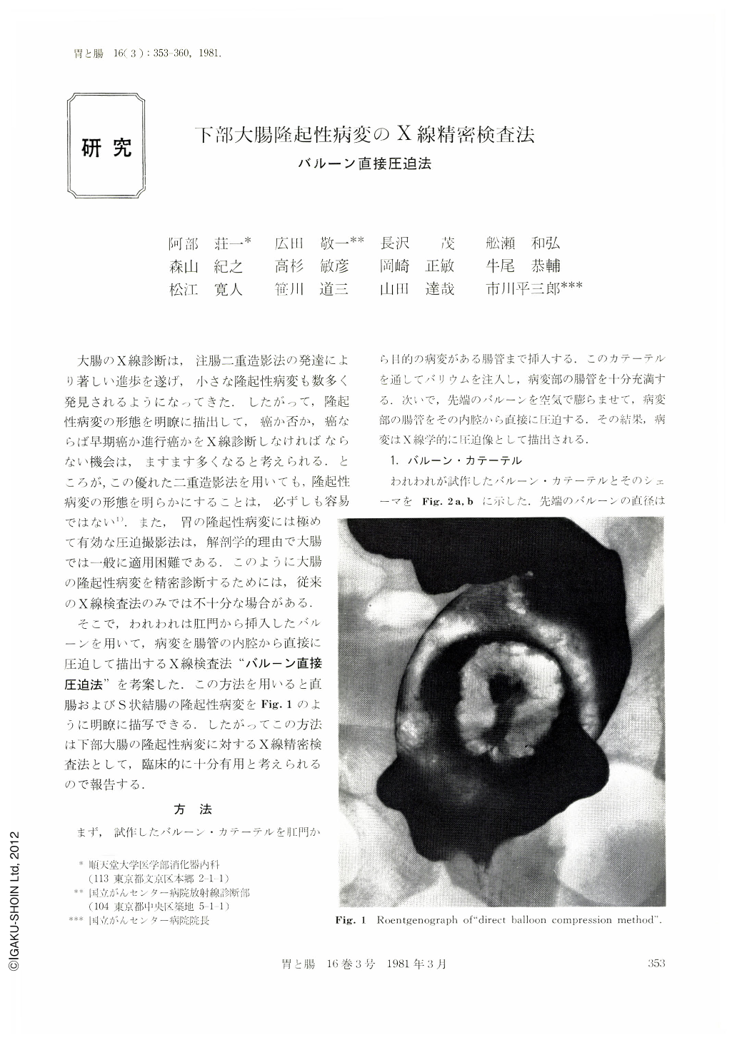
そこで,われわれは肛門から挿入したバルーンを用いて,病変を腸管の内腔から直接に圧迫して描出するX線検査法“バルーン直接圧迫法”を考案した.この方法を用いると直腸およびS状結腸の隆起性病変をFig.1のように明瞭に描写できる.したがってこの方法は下部大腸の隆起性病変に対するX線精密検査法として,臨床的に十分有用と考えられるので報告する.
Most of early cancers in the large bowel are usually polypoid types macroscopically. Recently, very small polypoid lesions in the large bowel can be detected rather easily with double contrast radiography. This technique, however, is not always successful in demonstrating detailed features of the polypoid lesion, especially in its central depression which is an important finding in making the diagnosis of cancer. In addition, it is almost impossible to employ the compression radiography in cases located at the rectum and sigmoid colon where most of cancer of the colon occur. Therefore, a new technique,“direct balloon compression method”, was developed and its usefulness to demonstrate detailed features of polypoid lesions was studied.
Whenever the polypoid lesions at the rectum and sigmoid colon were detected with double contrast examination, a balloon-catheter was inserted from the anus up to the region of the bowel where the polypoid lesions were found. After infusing 100~200ml of barium meal through the catheter, a balloon attached to the top of the catheter was inflated with air. The bowel where the polypoid lesion is located was then distended by the expanding balloon, which removed the excess barium and demonstrated detailed features of the polypoid lesion as an image of compression radiograph.
This method has been tried in various types of polypoid lesions including early cancers as well as Borrmann 2 type of advanced cancers. 23 lesions in the rectum and eight in the sigmoid colon were examined.
From the observation of these lesions, it was found that not only the central depressions of polypoid lesions but also their detailed features were demonstrated clearly with this “direct balloon compression method”. In conclusion, it shows that this new technique is applicable clinically, particularly for differential diagnosis of polypoid lesions which are located at the rectum and the sigmoid colon.

Copyright © 1981, Igaku-Shoin Ltd. All rights reserved.


