Japanese
English
- 有料閲覧
PPV購入案内
1,320円 (1,200円+税10%) こちらは、649ページから652ページの文献です。購入には医書.jp本会員登録が必要です。
会員登録完了後に改めて上記「購入サイトへ」を選択してください。
または「購入サイトへ」選択後に「購入ログイン」、「新規会員登録」の順に進んでください。
会員登録について詳しくはをご確認ください。
- Abstract 文献概要
- 1ページ目 Look Inside
はじめに
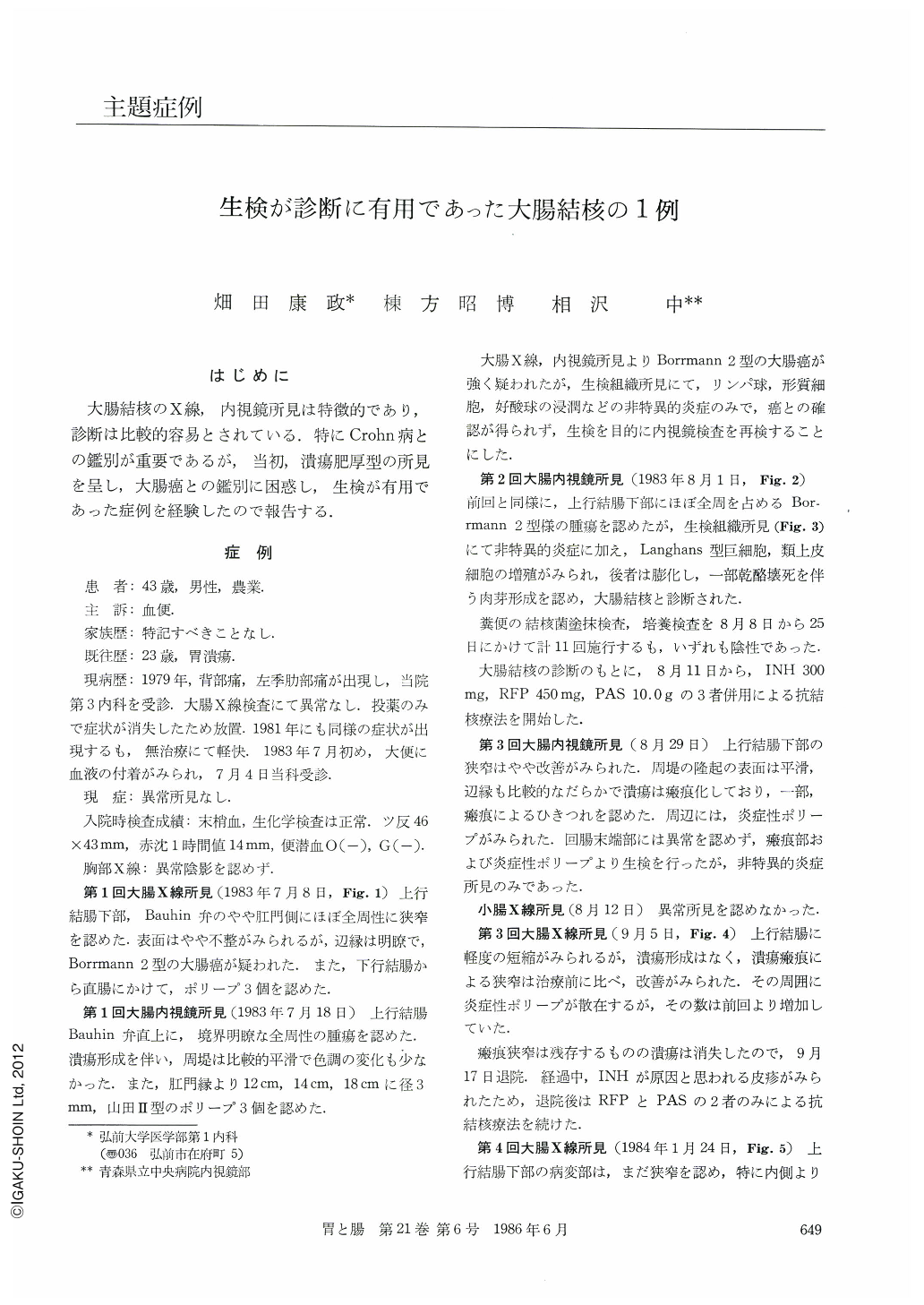
大腸結核のX線,内視鏡所見は特徴的であり,診断は比較的容易とされている.特にCrohn病との鑑別が重要であるが,当初,潰瘍肥厚型の所見を呈し,大腸癌との鑑別に困惑し,生検が有用であった症例を経験したので報告する.
A 43-year-old man was admitted to our hospital because of bloody stool. Physical examination was unremarkable. Laboratory studies revealed an erythrocyte sedimentation rate of 14mm/hr and tuberculin test yielded 46×43mm in erythelna. He had no history of pulmonary tuberculosis and chest roentogenogram was negative.
Barium enema x-ray study demonstrated annular stricture of the ascending colon with no shortening. Endoscopic examination showed a tumor with ulceration suggesting Borrmann type 2 colon cancer and three polyps in the rectosigmoid colon. Biopsy specimens, however, revealed caseating granuloma result-ing in the diagnosis of colonic tuberculosis. Repeated fecal cultures were all negative for acid-fast baccilli. The antituberculous drugs (INH, RFP & PAS) were administered.
After the treatment, annular stricture with ulceration of the ascending colon has improved into scars, followed by the formation of inflammatory polyps.
In this case it was difficult to differentiate colonic tuberculosis from cancer because of the segmental hypertrophic form and colonoscopic biopsy was useful in reaching the final diagnosis.
Copyright © 1986, Igaku-Shoin Ltd. All rights reserved.