Japanese
English
- 有料閲覧
- Abstract 文献概要
- 1ページ目 Look Inside
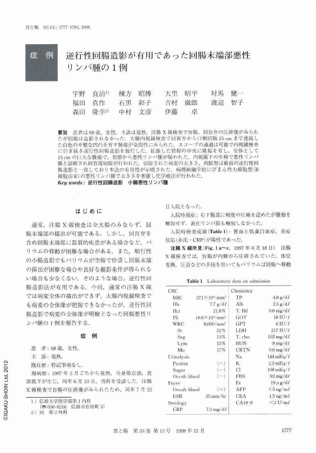
要旨 患者は68歳,女性.主訴は発熱.注腸X線検査で盲腸,回盲弁の圧排像がみられたが回腸は造影されなかった.大腸内視鏡検査で回盲弁から口側回腸15cmまで連続した白色の不整な凹凸を有す腫瘍が全周性にみられた.スコープの通過は可能で内視鏡検査に引き続き逆行性回腸造影を施行した.拡張した管腔の中央に隆起を有し,全体として15cmの巨大な腫瘍で,形態から悪性リンパ腫が疑われた.内視鏡下の生検で悪性リンパ腫と診断され回盲部切除が行われた.切除された病変の大きさ,肉眼型は術前の逆行性回腸造影と一致しており本法の有用性が示唆された.病理組織学的にびまん性大細胞型(B細胞由来)の悪性リンパ腫で大きさを考慮し化学療法が行われた.
A 68-year-old woman visited to our hospital because of fever. Barium enema revealed deformity of the ileocecal valve and cecum. However, the terminal portion of the ileum was not revealed. Colonoscopy showed a white-coated annular tumor with an irregular surface extending from the ileocecal valve to the proximal part of the ileum. Retrograde ileography was performed after colonoscopy, and this revealed a huge tumor (15 cm long) with a thick wall (4 cm). The tumor lumen was slightly widened and ragged. Malignant lymphoma was suspected. The histologic diagnosis obtained by endoscopic biopsy specimen was malignant lymphoma of the diffuse large cell type. The patient underwent right hemicolectomy and chemotherapy. It has been reported that retrograde ileography is useful for detection of lesions in the terminal portion of the ileum, and this was a typical example of such a case.

Copyright © 1998, Igaku-Shoin Ltd. All rights reserved.


