Japanese
English
- 有料閲覧
- Abstract 文献概要
- 1ページ目 Look Inside
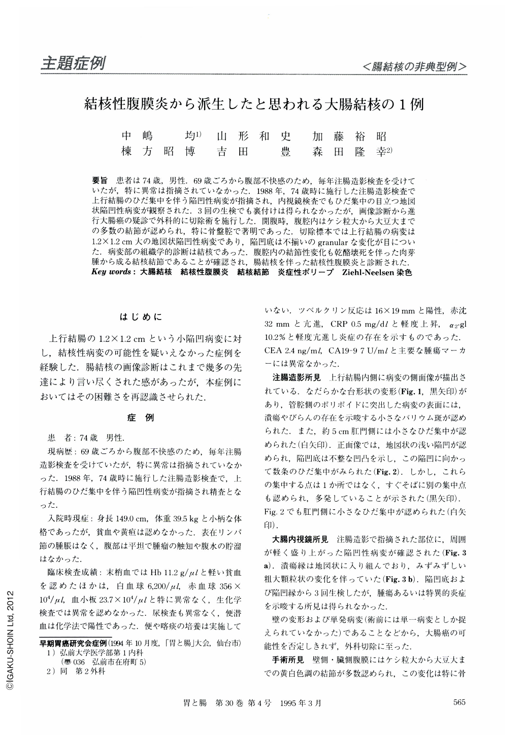
要旨 患者は74歳,男性.69歳ごろから腹部不快感のため,毎年注腸造影検査を受けていたが,特に異常は指摘されていなかった.1988年,74歳時に施行した注腸造影検査で上行結腸のひだ集中を伴う陥凹性病変が指摘され,内視鏡検査でもひだ集中の目立つ地図状陥凹性病変が観察された.3回の生検でも裏付けは得られなかったが,画像診断から進行大腸癌の疑診で外科的に切除術を施行した.開腹時,腹腔内はケシ粒大から大豆大までの多数の結節が認められ,特に骨盤腔で著明であった.切除標本では上行結腸の病変は1.2×1.2cm大の地図状陥凹性病変であり,陥凹底は不揃いのgranularな変化が目についた.病変部の組織学的診断は結核であった.腹腔内の結節性変化も乾酪壊死を伴った肉芽腫から成る結核結節であることが確認され,腸結核を伴った結核性腹膜炎と診断された.
A 74-year-old man was hospitalized with the complaint of abdominal discomfort. Barium enema examination and colonoscopy revealed a geographically ulcerated lesion with remarkable fold convergence in the ascending colon. We repeated biopsies three times, but specific findings including malignancy could not be obtained from those specimens. We couldn't deny the possibility of colon cancer, and carried out right hemicolectomy because we strongly suspected colon cancer. Laparotomy showed multiple small nodule sized from 1~6 mm all over the peritoneal cavity. The changes in the pelvis were remarkable, but ascites mas not found. We found a 1.2 × 1.2 cm sized ulcerated lesion in the middle of the ascending colon. Histological examination of the resected specimen proved it to be colon tuberculosis accompanied by peritonitis tuberculosa.

Copyright © 1995, Igaku-Shoin Ltd. All rights reserved.


