Japanese
English
- 有料閲覧
PPV購入案内
1,320円 (1,200円+税10%) こちらは、1109ページから1112ページの文献です。購入には医書.jp本会員登録が必要です。
会員登録完了後に改めて上記「購入サイトへ」を選択してください。
または「購入サイトへ」選択後に「購入ログイン」、「新規会員登録」の順に進んでください。
会員登録について詳しくはをご確認ください。
- Abstract 文献概要
- 1ページ目 Look Inside
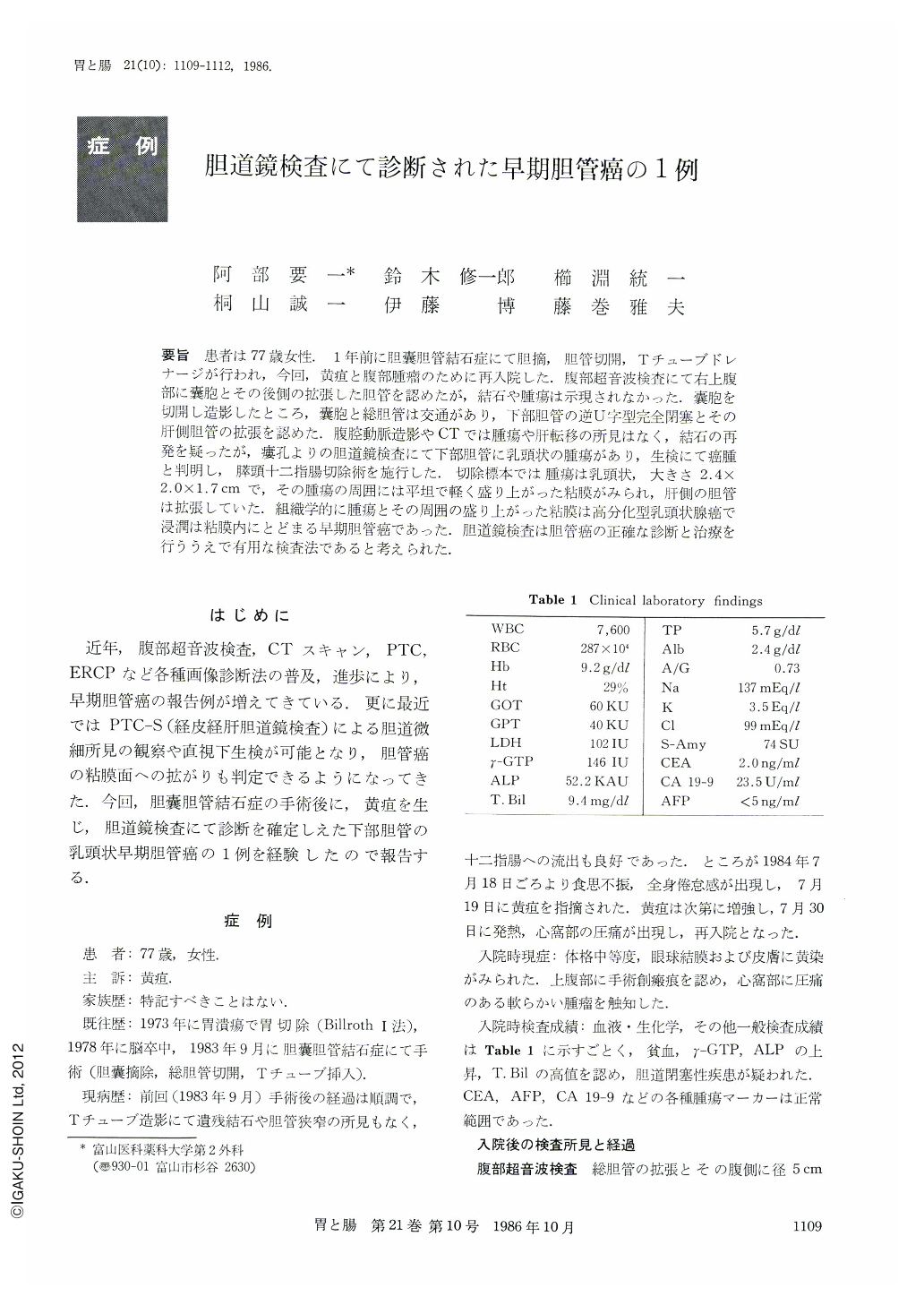
要旨 患者は77歳女性.1年前に胆嚢胆管結石症にて胆摘,胆管切開,Tチューブドレナージが行われ,今回,黄疸と腹部腫瘤のために再入院した.腹部超音波検査にて右上腹部に囊胞とその後側の拡張した胆管を認めたが,結石や腫瘍は示現されなかった.囊胞を切開し造影したところ,囊胞と総胆管は交通があり,下部胆管の逆U字型完全閉塞とその肝側胆管の拡張を認めた.腹腔動脈造影やCTでは腫瘍や肝転移の所見はなく,結石の再発を疑ったが,痕孔よりの胆道鏡検査にて下部胆管に乳頭状の腫瘍があり,生検にて癌腫と判明し,膵頭十二指腸切除術を施行した.切除標本では腫瘍は乳頭状,大きさ2.4×2.0×1.7cmで,その腫瘍の周囲には平坦で軽く盛り上がった粘膜がみられ,肝側の胆管は拡張していた.組織学的に腫瘍とその周囲の盛り上がった粘膜は高分化型乳頭状腺癌で浸潤は粘膜内にとどまる早期胆管癌であった.胆道鏡検査は胆管癌の正確な診断と治療を行ううえで有用な検査法であると考えられた.
The patient, a 77 year-old woman, received cholecystectomy, choledochotomy and T-tube drainage because of cholecystocholedocholithiasis. After a year, she was admitted to our hospital with complaints of jaundice and abdominal tumor. Abdominal US examination revealed a cyst at the right upper abdomen and dilatation of the choledochus behind the cyst. But neither stone nor tumor was shown in the echo.
The cyst was incised. Cystography revealed a fistula between the cyst and the choledochus.
Cholangiogram demonstrated completely reversed U-shaped obstruction of the distal bile duct and marked dilatation of the common and intrahepatic bile duct. Celiac angiogram and CT scan did not show the tumor and liver metastasis. Therefore we suspected recurrence of a stone. The cholangioscope was inserted into the choledochus through the fistula. Cholangioscopic finding revealed a papillomatous tumor in the distal bile duct. Histologically, the tumor was a carcinoma. Pancreatoduodenectomy was performed. Operative finding showed H0, P0, S0, N (-), Hinf0, G0, Panc0, D0, V0, that is Stage Ⅰ, so an absolute curative resection was carried out. Resected specimen showed the tumor was 2.4×2.0×1.7 cm in size and papillomatous.
The mucosa around the tumor was slightly elevated. Histologically the tumor and the elevated mucosa were well differentiated papillary adenocarcinoma located in the mucosal layer of the bile duct.
We consider that cholangioscopy may be useful for the diagnosis and treatment of a bile duct tumor.
Copyright © 1986, Igaku-Shoin Ltd. All rights reserved.