Japanese
English
- 有料閲覧
PPV購入案内
1,320円 (1,200円+税10%) こちらは、705ページから713ページの文献です。購入には医書.jp本会員登録が必要です。
会員登録完了後に改めて上記「購入サイトへ」を選択してください。
または「購入サイトへ」選択後に「購入ログイン」、「新規会員登録」の順に進んでください。
会員登録について詳しくはをご確認ください。
- Abstract 文献概要
- 1ページ目 Look Inside
- サイト内被引用 Cited by
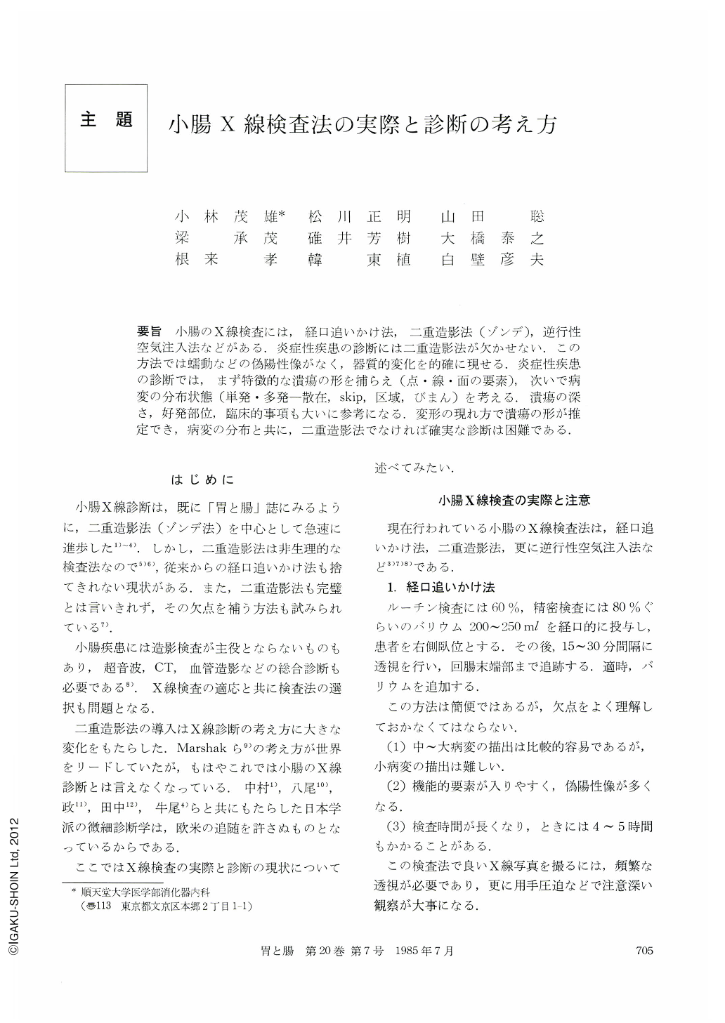
要旨 小腸のX線検査には,経口追いかけ法,二重造影法(ゾンデ),逆行性空気注入法などがある.炎症性疾患の診断には二重造影法が欠かせない.この方法では蠕動などの偽陽性像がなく,器質的変化を的確に現せる.炎症性疾患の診断では,まず特徴的な潰瘍の形を捕らえ(点・線・面の要素),次いで病変の分布状態(単発・多発-散在,skip,区域,びまん)を考える.潰瘍の深さ,好発部位,臨床的事項も大いに参考になる.変形の現れ方で潰瘍の形が推定でき,病変の分布と共に,二重造影法でなければ確実な診断は困難である.
The double contrast method by means of duodenal intubation and administration of barium and air is best suited for diagnosis of the small bowel disease.
Merits of this method are as follows.
1) Examination time is shorter than follow through method.
2) False positive findings by peristalsis can eliminated by injection of antispasmodics.
3) Not only big but small and fine mucosal abnormalities can be well observed.
The shapes of ulcerations of inflammatory bowel disease are divided to Point, Line and Area and for presumption of them, deformity of the wall is the most important indication to be looked for.
The distribution pattern of ulcerative lesions is Single or multiple-Sporadic, Segmental or Diffuse.
Characteristic findings can be found in each inflammatory disease by analysis of the shape of ulcerations and the distribution pattern.
Inflammatory small bowel disease should be diagnosed by double contrast method by studying the shape and the distribution pattern of ulcerative lesions.
Copyright © 1985, Igaku-Shoin Ltd. All rights reserved.