Japanese
English
- 有料閲覧
- Abstract 文献概要
- 1ページ目 Look Inside
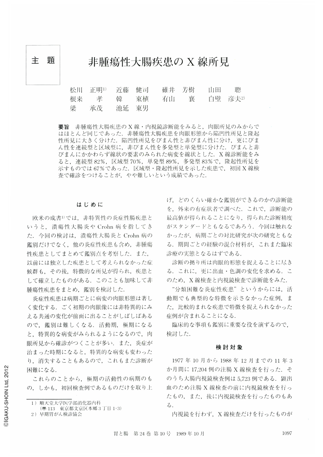
要旨 非腫瘍性大腸疾患のX線・内視鏡診断能をみると,肉眼所見のみからではほとんど同じであった.非腫瘍性大腸疾患を肉眼形態から陥凹性所見と隆起性所見に大きく分けた.陥凹性所見をびまん性と非びまん性に分け,更にびまん性を連続型と区域型に,非びまん性を多発型と単発型に分けた.びまんと非びまんにかかわらず線状の要素のみられた病変を線状とした.X線診断能をみると,連続型82%,区域型70%,単発型89%,多発型83%で,隆起性所見を示すものでは67%であった.区域型・隆起性所見を示した疾患で,初回X線検査で確診をつけることが,やや難しいという成績であった.
Study was conducted regarding differential diagnosis of non-neoplastic diseases using radiological findings in 225 cases (UC 117, ischemic colitis 26, drug-induced colitis 23, Crohn's disease 21, so-called simple ulcer 20, intestinal tuberculosis 3, amyloidosis 3, amebiasis 3, others 9 cases).
Classification was possible radiologically as follows; diffusely depressed form (diffuse form), non-diffusely depressed form (non-diffuse form) and protruded form. Diffuse form then consists of continuous and segmental types. Continuous type describes diffuse ulcerative lesion proximal to the rectum or of the rectum. While segmental type represents a diffuse ulcerative lesion in one part of the colon except the rectum. Non-diffuse form may radiologically represent normal mucosa scattered among the ulcerative lesions. Non-diffuse form consists of multiple and simple types.
Application of our radiological classification yielded correct diagnosis in 87% for continuous type, 69% for segmental type, 89% for simple type, 83% for multiple type and 67% for protruded form. Making a diagnosis of non-neoplastic colorectal diseases was thus difficult for the segmental type followed by the protruded form.

Copyright © 1989, Igaku-Shoin Ltd. All rights reserved.


