Japanese
English
- 有料閲覧
- Abstract 文献概要
- 1ページ目 Look Inside
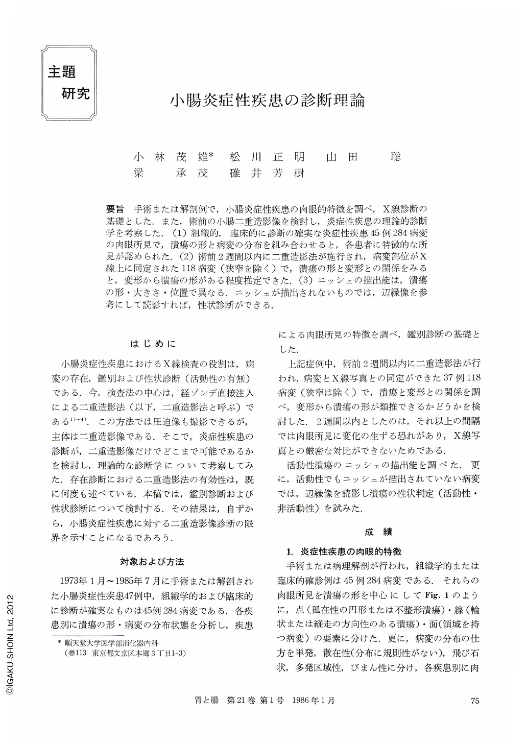
要旨 手術または解剖例で,小腸炎症性疾患の肉眼的特徴を調べ,X線診断の基礎とした.また,術前の小腸二重造影像を検討し,炎症性疾患の理論的診断学を考察した.(1)組織的,臨床的に診断の確実な炎症性疾患45例284病変の肉眼所見で,潰瘍の形と病変の分布を組み合わせると,各患者に特徴的な所見が認められた.(2)術前2週間以内に二重造影法が施行され,病変部位がX線上に同定された118病変(狭窄を除く)で,潰瘍の形と変形との関係をみると,変形から潰瘍の形がある程度推定できた.(3)ニッシェの描出能は,潰瘍の形・大きさ・位置で異なる.ニッシェが描出されないものでは,辺縁像を参考にして読影すれば,性状診断ができる.
(1) Forty-five cases (284 lesions) of inflammatory small bowel disease were operated or dissected in Juntendo University Hospital during 13 years from 1973 to 1985. For the basis of x-ray diagnosis, macroscopic appearance was analized by shape of the ulceration and distribution pattern. Ulcerations are devided into three groups by shape of them, Point, Line and Area. Distribution pattern of the lesions was recognized as single or multiplicity which was classified to sporadic, skip, segmental or diffuse. Characteristic macroscopic findings (shape of ulceration and distribution pattern) could be found in each inflammatory disease. Double contrast method was the best suited for differential diagnosis of inflammatory disease because it had excellent demonstrability of ulcerative lesions.
(2) Double contrast method by means of duodenal intubation was done in 30 cases within two weeks before operation. Deformity of the wall was closely related with shape, size and position of the ulceration. Shape of ulceration couldbe estimated by the analysis of deformity of the wall when the niche could not be demonstrated.
(3) Irregularity of the wall was very helpful for diagnosis of whether active or inactive ulcerative lesions. Almost of active stage ulcerations except small size had irregulality of the wall and some inactive stage ulceraton had the same finding. Diagnosis of properties of ulceration could be made by demonstration of niche and irregularity of the wall.

Copyright © 1986, Igaku-Shoin Ltd. All rights reserved.


