Japanese
English
- 有料閲覧
- Abstract 文献概要
- 1ページ目 Look Inside
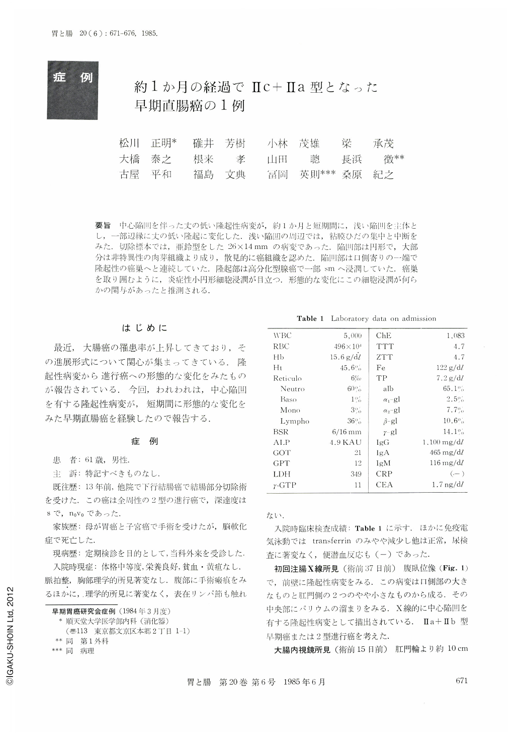
要旨 中心陥凹を伴った丈の低い隆起性病変が,約1か月と短期間に,浅い陥凹を主体とし,一部辺縁に丈の低い隆起に変化した.浅い陥凹の周辺では,粘膜ひだの集中と中断をみた,切除標本では,亜鈴型をした26×14mmの病変であった.陥凹部は円形で,大部分は非特異性の肉芽組織より成り,散見的に癌組織を認めた.陥凹部は口側寄りの一端で隆起性の癌巣へと連続していた.隆起部は高分化型腺癌で一部smへ浸潤していた.癌巣を取り囲むように,炎症性小円形細胞浸潤が目立つ.形態的な変化にこの細胞浸潤が何らかの関与があったと推測される.
Most of early colorectal cancer cases reported were of the protruded types, and the depressed types have been reported rarely. Our case was demonstrated by radiography as a protruded rectal lesion that transformed into Ⅱc+Ⅱa type early rectal cancer. The patients was a 61-year-old man without a chief complaint. Thirteen years before this admission, he had been operated on in order to resect an advanced cancer of the colon descendens. Thirty-seven days before this operation, a nodular lesion with a central depression was radiologically demonstrated at the rectum. Fifteen days before this, a fine granular plaque-like lesion was observed by endoscope at oral side of the lesion and easily fragile mucosa at its anal side. Three days before this, the rectal lesion consisted of one part of a shallow depression and the other of a plaque-like lesion. Some converging folds around the depressed part were seen. Close-up view of the resected specimen, a dumbbell-shaped lesion measuring 26×14mm in diameter, consisted of flattened vinous elevation and a shallow round depreslion. Histologically, the lesion disclosed infiltration of well differentiated adenocarcinoma in the submucosa without any evidence of an adenoma.

Copyright © 1985, Igaku-Shoin Ltd. All rights reserved.


