Japanese
English
- 有料閲覧
PPV購入案内
1,320円 (1,200円+税10%) こちらは、637ページから640ページの文献です。購入には医書.jp本会員登録が必要です。
会員登録完了後に改めて上記「購入サイトへ」を選択してください。
または「購入サイトへ」選択後に「購入ログイン」、「新規会員登録」の順に進んでください。
会員登録について詳しくはをご確認ください。
- Abstract 文献概要
- 1ページ目 Look Inside
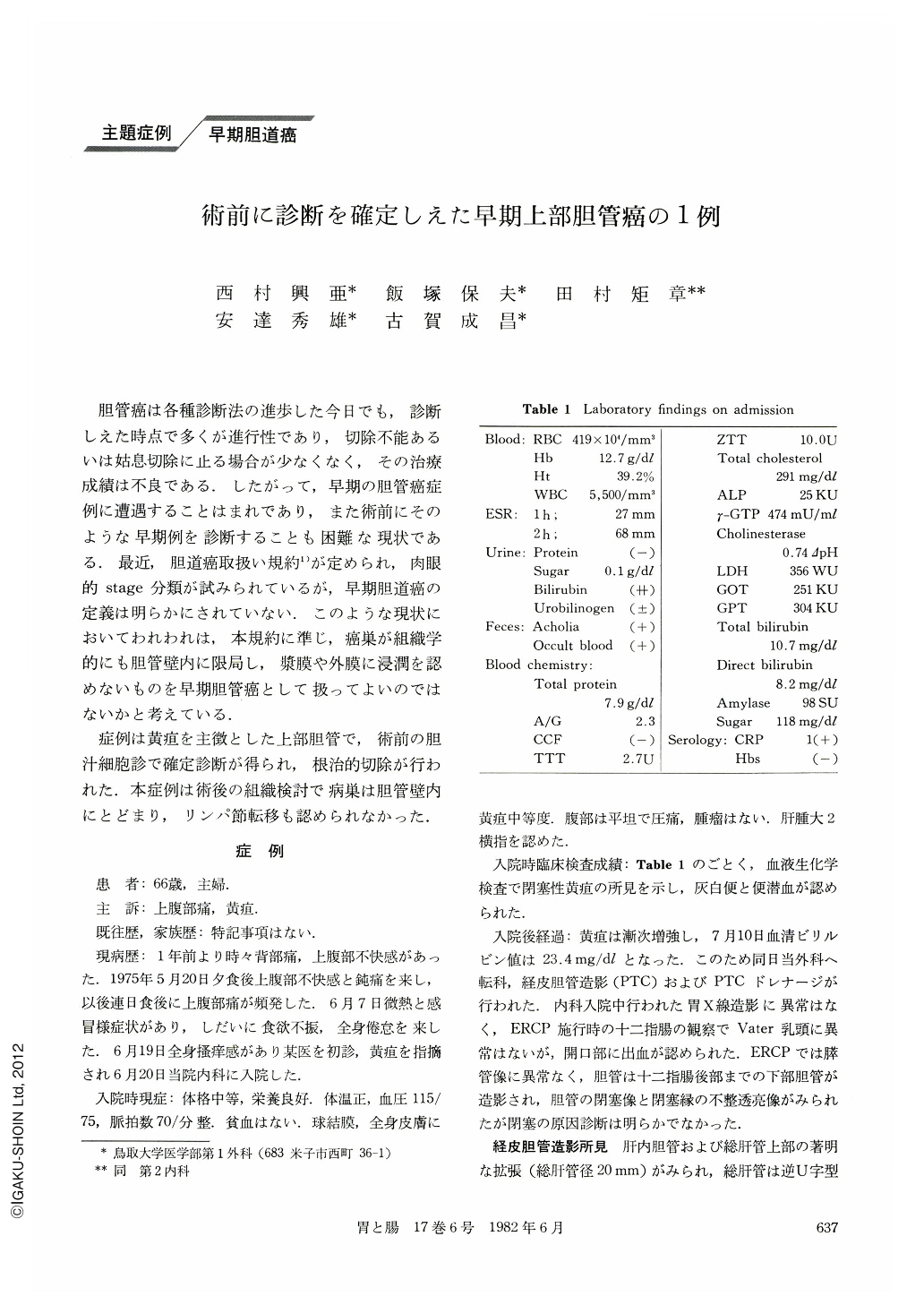
胆管癌は各種診断法の進歩した今日でも,診断しえた時点で多くが進行性であり,切除不能あるいは姑息切除に止る場合が少なくなく,その治療成績は不良である.したがって,早期の胆管癌症例に遭遇することはまれであり,また術前にそのような早期例を診断することも困難な現状である.最近,胆道癌取扱い規約1)が定められ,肉眼的stage分類が試みられているが,早期胆道癌の定義は明らかにされていない.このような現状においてわれわれは,本規約に準じ,癌巣が組織学的にも胆管壁内に限局し,漿膜や外膜に浸潤を認めないものを早期胆管癌として扱ってよいのではないかと考えている.
症例は黄疸を主徴とした上部胆管で,術前の胆汁細胞診で確定診断が得られ,根治的切除が行われた.本症例は術後の組織検討で病巣は胆管壁内にとどまり,リンパ節転移も認められなかった.
A 66-year-old woman was admitted to our hospital on June 20, 1978, because of epigastric pain and jaundice. Laboratory examinations on admission revealed obstructive jaundice with a level of 10.7 mg/dl of serum total bilirubin.
Percutaneous transhepatic cholangiography (PTC) showed marked dilatation of bile ducts and an obstructive shadow with reversed U sign in the common hepatic duct, as if it were an incarcerated stone. But, the obstruction was incomplete, and it was suggested as a tumor lesion because of its marginal appearance, presenting a shadow defect, or by lack of mobility.
Cytologic examination which was studied by biliary washing technique via PTC drainage revealed some malignant cells. And the lesion was confirmed preoperatively as carcinoma of the common hepatic duct.
The patient was operated on on July 28, 18 days after biliary decompression by PTC drainage. An index finger's head sized tumor was found in the common hepatic duct on laparotomy, and neither metastatic lesions nor local invasions were observed. So, the radical resection of the extrahepatic bile duct with regional lymph nodes dissection was carried out, and hepaticojejunostomy by Roux en Y was performed. On exploration of the resected specimen, a limited nodular type tumor measuring 2.7×1.4×1.1 cm was noticed in the left sided wall of the bile duct. Histological study revealed moderately differentiated tubular adenocarcinoma, partly involving papillary or mucinous adenocarcinoma. The lesion infiltrated the duct wall, but limited without serosal or adventitial invasions. Neither vascular nor perineural invasion could be identified. And, the dissected lymph nodes examined were negative for cancer cells.
The patient's postoperative course was uneventful and she was discharged on the 16th postoperative day. And 41 months later, she is alive and well.
Copyright © 1982, Igaku-Shoin Ltd. All rights reserved.