Japanese
English
- 有料閲覧
PPV購入案内
1,320円 (1,200円+税10%) こちらは、889ページから894ページの文献です。購入には医書.jp本会員登録が必要です。
会員登録完了後に改めて上記「購入サイトへ」を選択してください。
または「購入サイトへ」選択後に「購入ログイン」、「新規会員登録」の順に進んでください。
会員登録について詳しくはをご確認ください。
- Abstract 文献概要
- 1ページ目 Look Inside
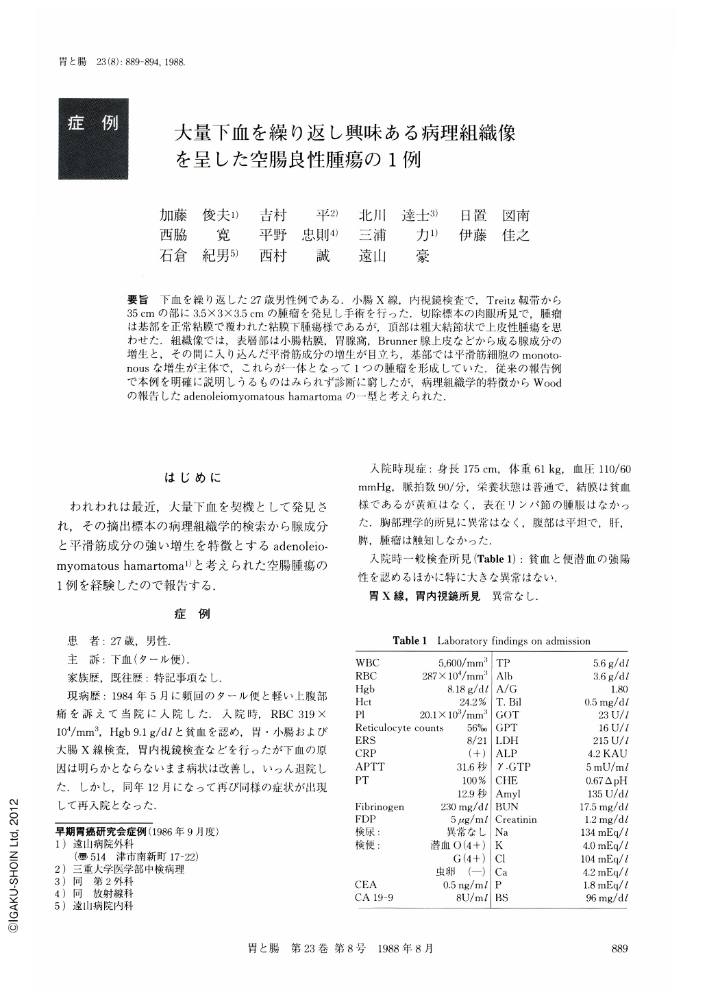
要旨 下血を繰り返した27歳男性例である.小腸X線,内視鏡検査で,Treitz靱帯から35cmの部に3.5×3×3.5cmの腫瘤を発見し手術を行った.切除標本の肉眼所見で,腫瘤は基部を正常粘膜で覆われた粘膜下腫瘍様であるが,頂部は粗大結節状で上皮性腫瘍を思わせた.組織像では,表層部は小腸粘膜,胃腺窩,Brunner腺上皮などから成る腺成分の増生と,その間に入り込んだ平滑筋成分の増生が目立ち,基部では平滑筋細胞のmonotonousな増生が主体で,これらが一体となって1つの腫瘤を形成していた.従来の報告例で本例を明確に説明しうるものはみられず診断に窮したが,病理組織学的特徴からWoodの報告したadenoleiomyomatous hamartomaの一型と考えられた.
A 27 year-old man presented with repeated attacks of melena. A barium study and an endoscopic study revealed a mass lesion within the jejunum 30 to 40 cm anal to the ligament of Treitz (Figs. 1 and 2). The mass was resected with 30 cm of the adjacent jejunum.
Gross examination of the resected specimen disclosed the mass, measuring 3.5×3.0×3.5 cm, with the base being covered with normal-looking intestinal mucosa, and the nodular top being covered with fine granular epithelium (Fig. 3). Histopathological findings were characteristic in that there were proliferation of glandular cells as well as smooth muscle fibers in the luminal surface (Fig. 5) and leiomyoma-like proliferation of smooth muscle fibers in the base (Fig. 6). The glandular elements consisted of small intestinal mucosa, foveolar epithelium of the stomach and Brunner's gland, and there was no cellular atypia.
There has been no reports of jejunal tumor showing such characteristic histopathological findings as in this case. We consider, however, that this case is probably a variant form of adenoleiomyomatous hamartoma described by Wood.
Copyright © 1988, Igaku-Shoin Ltd. All rights reserved.