Japanese
English
- 有料閲覧
PPV購入案内
1,320円 (1,200円+税10%) こちらは、1061ページから1066ページの文献です。購入には医書.jp本会員登録が必要です。
会員登録完了後に改めて上記「購入サイトへ」を選択してください。
または「購入サイトへ」選択後に「購入ログイン」、「新規会員登録」の順に進んでください。
会員登録について詳しくはをご確認ください。
- Abstract 文献概要
- 1ページ目 Look Inside
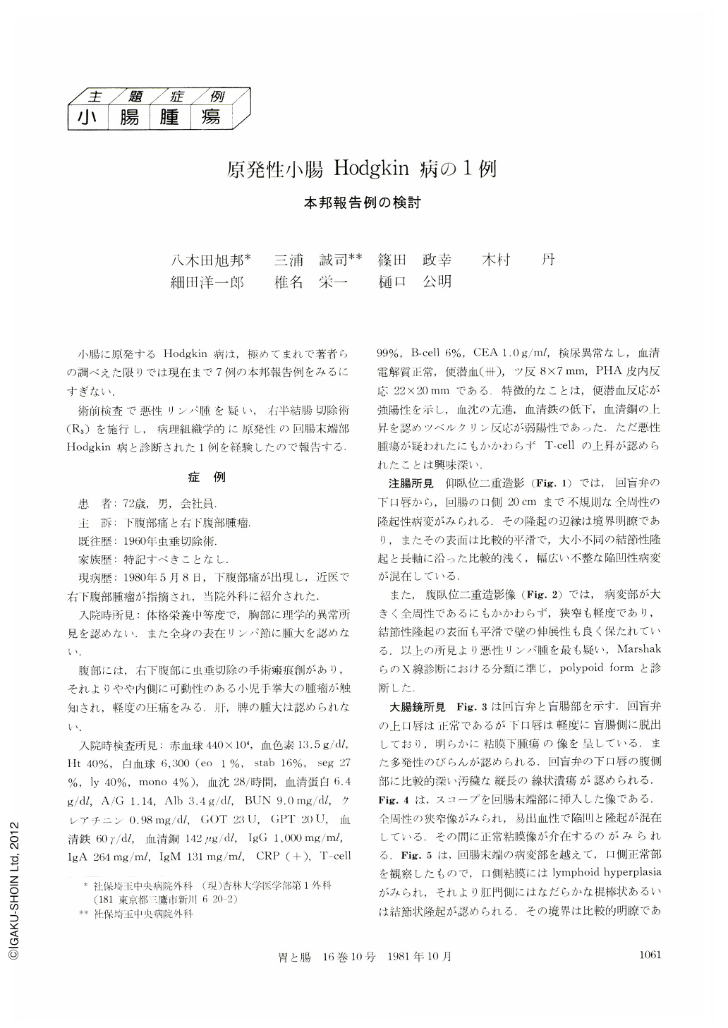
小腸に原発するHodgkin病は,極めてまれで著者らの調べえた限りでは現在まで7例の本邦報告例をみるにすぎない.
術前検査で悪性リンパ腫を疑い,右半結腸切除術(R3)を施行し,病理組織学的に原発性の回腸末端部Hodgkin病と診断された1例を経験したので報告する.
A 72-year-old man visited the Saitama Chuo hospital on, July 15, 1980, with the chief complaints of palpable mass at the right lower quadrant and lower abdominal pain, which had started since May, 1980. In laboratoy findings, the feces was positive for occult blood.
A supine double contrast view revealed a tumortype lesion with shallow irregular depressions and several nodes of smooth margin at ileum end. Prone double contract film demonstrated a smooth and large polypoid lesion at the oral site of the ileo-cecal valve. Endoscopic picture revealed a submucosal tumor with multiple erosions and linear ulcer at the under lip of the ileo-cecal valve, which is prolapsed to the cecum. Colonofiberscopic picture showed a narrow segment because of protuberant growth lesion with irregular ulceration of the surface at the ileum end. Biopsy performed twice was negative for malignancy. Operation was performed on July 30, 1980 with a suspected diagnosis of malignant lymphoma. Right hemicolectomy-curative operative (R3) was done (N1 H0 P0 S2).
In the resected ileum end and right side colon, there was an elastic and hard gyros-like tumor of the ileum end with erosions and shallow ulcerations, socalled the protuberant growth, measuring about 85×75X30mm, which was well demarcated. The tumor cell infilerated into the serosa and two out of eight regional lymph nodes were invaded. According to Rye's classification of Hodgkin's disease, the tumor belongs histologically to the lymphocytic depletion type.
Copyright © 1981, Igaku-Shoin Ltd. All rights reserved.