Japanese
English
- 有料閲覧
PPV購入案内
1,320円 (1,200円+税10%) こちらは、919ページから926ページの文献です。購入には医書.jp本会員登録が必要です。
会員登録完了後に改めて上記「購入サイトへ」を選択してください。
または「購入サイトへ」選択後に「購入ログイン」、「新規会員登録」の順に進んでください。
会員登録について詳しくはをご確認ください。
- Abstract 文献概要
- 1ページ目 Look Inside
- サイト内被引用 Cited by
1963年にDe La Pavaら1)が,食道粘膜上皮基底層にmelanoblastsが存在することを証明して以来,食道原発性悪性黒色腫の存在が認識されてきたが,その頻度は極めてまれである.
今回,われわれは食道原発の悪性黒色腫の1例を経験したので,経過の概略を述べると共に,自験例を含め本邦報告例32例につき若干の文献的考察を行ったので報告する.
A case of primary malignant melanoma of the esophagus in a 35-year-old woman is reported.
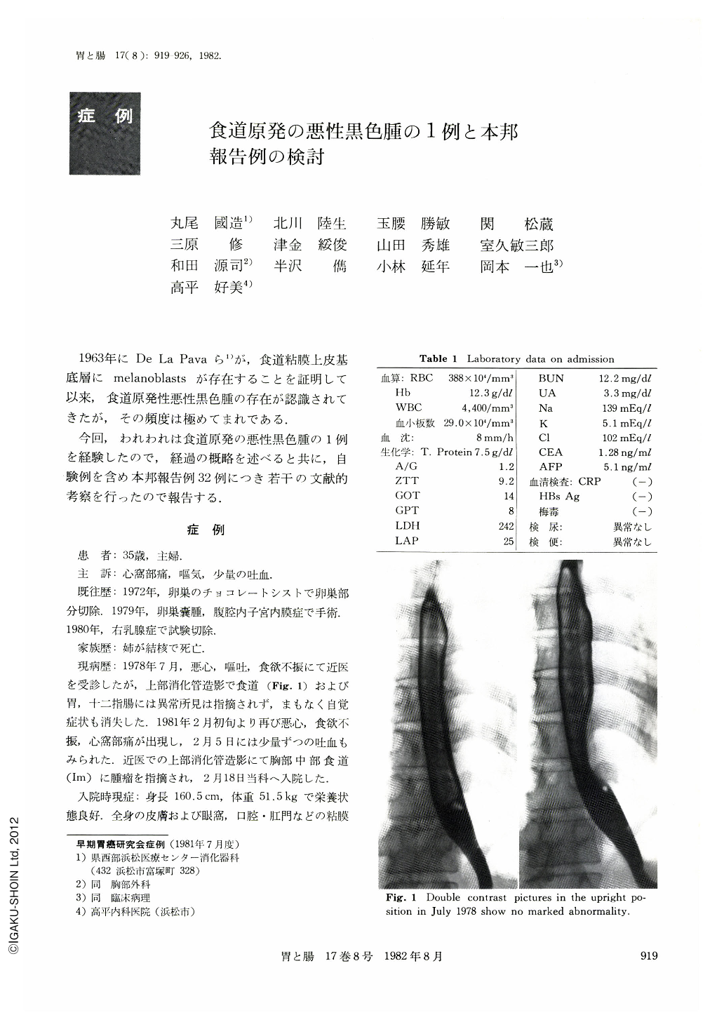
She was referred to Hamamatsu Medical Center in February 1981, complaining of epigastralgia, nausea and hematemesis for a few days duration. Clinical examination was unremarkable with no evidence of cutaneous, retinal or other melanotic lesions. A barium swallow study revealed a semipedunculated or pedunculated lesion with nodular surface in the middle thoracic esophagus and small elevated lesion 2 cm above the esophago-gastric junction. Esophagoscopy disclosed a semipedunculated tumor ; its surface was relatively smooth, partially nodulated with grape color appearance. And several small black elevated lesions were noted at a distance of 38 cm from the incisor teeth. Considering hemangiomas, no biopsy was carried out. On March 25, 1981, resections of tumors were performed. Examination of the two specimens disclosed relatively smooth masses, but partially lobulated with dark-bluish color measuring 2.9×2.9cm and 0.9×0.9cm respectively. Histologically, the large tumor showed extension into submucosa and muscle layers. They consisted mainly of epitheloid cells. Melanin pigment was frequently present in the tumor cells. Junctional activities were found in the adjacent squamous epithelium on both tumors. No lymph node metastasis was identified. After the operation, she was given 4,000 rad of 60Co irradiation. Though her course was uneventful, endoscopic study on July 28, 1981, showed a few blueblack spots on the esophageal mucosa. Biopsy proved atypical melanocytes. Against the spots, injections of BCG had been done every two weeks since August 11, 1981. Subjectively, she was well and no remarkable metastasis was seen till September 20, 1981.
A review of the literature on primary malignant melanoma of the esophagus enabled us to find a total of 32 cases, including our case, from 1960 to 1981 in Japan.
The commonest initial symptom was mild to moderate swallowing disturbance. The mean duration of symptoms was 3.8 months. The age distribution of 32 cases were 35 to 77 years. The ratio of male to female was 22: 10. Majority of cases were located in the middle or the lower third esophagus. They were pedunculated, polypoid or lobulated in appearance. The prognosis was always grave, for wide speed metastases developed. The greater part of the patients died within one year since the initial symptoms.
Therefore, detection of the disease at an early state while it is resectable seems to offer hope of curing it.
Copyright © 1982, Igaku-Shoin Ltd. All rights reserved.