Japanese
English
- 有料閲覧
PPV購入案内
1,320円 (1,200円+税10%) こちらは、223ページから227ページの文献です。購入には医書.jp本会員登録が必要です。
会員登録完了後に改めて上記「購入サイトへ」を選択してください。
または「購入サイトへ」選択後に「購入ログイン」、「新規会員登録」の順に進んでください。
会員登録について詳しくはをご確認ください。
- Abstract 文献概要
- 1ページ目 Look Inside
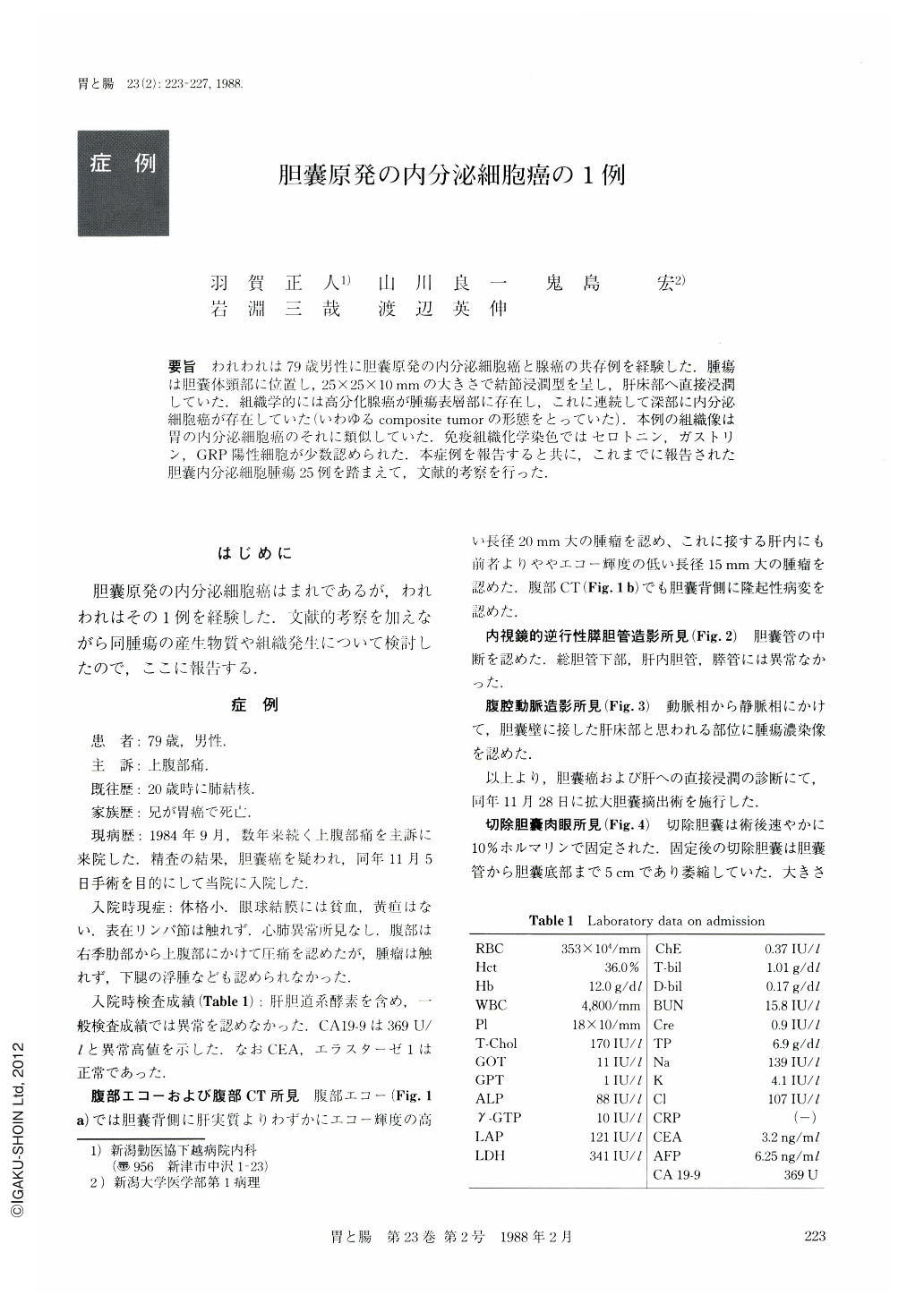
要旨 われわれは79歳男性に胆囊原発の内分泌細胞癌と腺癌の共存例を経験した.腫瘍は胆囊体頸部に位置し,25×25×10mmの大きさで結節浸潤型を呈し,肝床部へ直接浸潤していた.組織学的には高分化腺癌が腫瘍表層部に存在し,これに連続して深部に内分泌細胞癌が存在していた(いわゆるcomposite tumorの形態をとっていた).本例の組織像は胃の内分泌細胞癌のそれに類似していた.免疫組織化学染色ではセロトニン,ガストリン,GRP陽性細胞が少数認められた.本症例を報告すると共に,これまでに報告された胆囊内分泌細胞腫瘍25例を踏まえて,文献的考察を行った.
The patient was a 79 year-old man and was admitted to our hospital complaining of epigastric pain. Ultrasonographic and radiographic examination led us to establishing a diagnosis of the gallbladder carcimoma. He underwent extended cholecystectomy.
Macroscopically, the resected gallbladder was small, but revealed a nodular infiltraing tumor measuring 25×25×15 mm. Microscopically, the tumor was composed of papillotubular adenocarcinoma and endocrine cell carcinoma. The latter had invaded the liver bed directly. Tumor cells had round vesicular nuclei, and scanty cytoplasm with positive granules shown by Grimelius' method. The endocrine cell carcinoma showed several serotonin, GRP, and gastrin-positive cells shown by PAP method.
We know only a small number of reports of primary endocrine cell tumor of the gallbladder, 25 cases as far as we could trace. The purpose of this paper is to describe clinical and histological characteristics of the endocrine cell carcinoma. We must distinguish endocrine cell carcinoma from the classical carcinoid on the points of cellular atypia, pleomorphism, mitotic figures, vascular permeation, metastasis in its early stage, and poor prognosis.
Copyright © 1988, Igaku-Shoin Ltd. All rights reserved.