Japanese
English
- 有料閲覧
PPV購入案内
1,320円 (1,200円+税10%) こちらは、253ページから261ページの文献です。購入には医書.jp本会員登録が必要です。
会員登録完了後に改めて上記「購入サイトへ」を選択してください。
または「購入サイトへ」選択後に「購入ログイン」、「新規会員登録」の順に進んでください。
会員登録について詳しくはをご確認ください。
- Abstract 文献概要
- 1ページ目 Look Inside
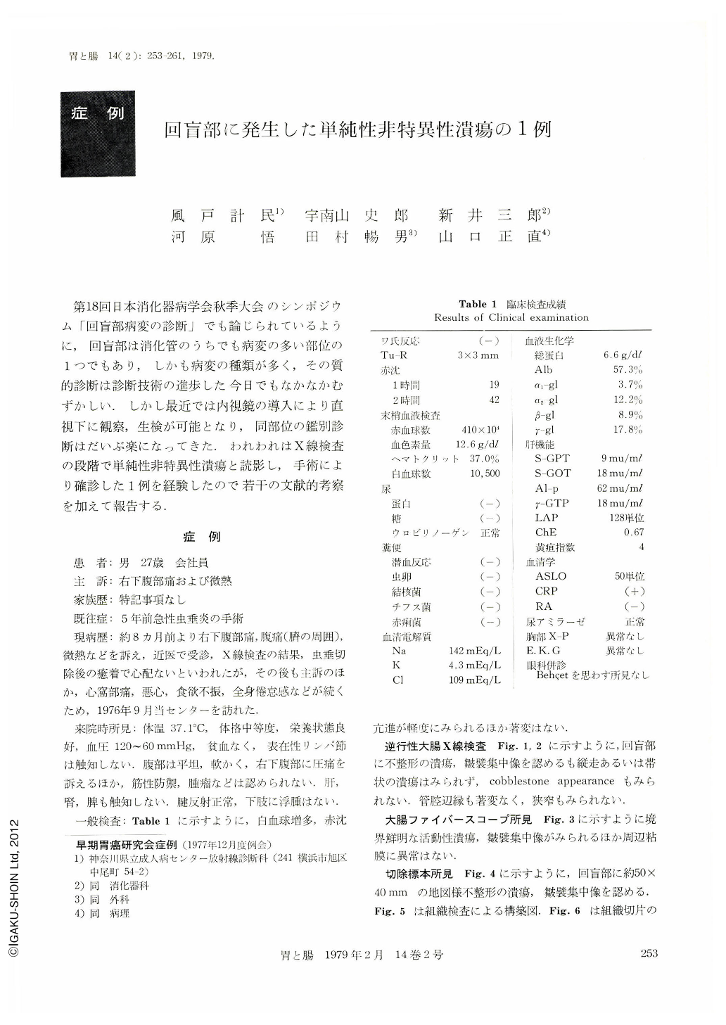
第18回日本消化器病学会秋季大会のシンポジウム「回盲部病変の診断」でも論じられているように,回盲部は消化管のうちでも病変の多い部位の1つでもあり,しかも病変の種類が多く,その質的診断は診断技術の進歩した今日でもなかなかむずかしい.しかし最近では内視鏡の導入により直視下に観察,生検が可能となり,同部位の鑑別診断はだいぶ楽になってきた.われわれはX線検査の段階で単純性非特異性潰瘍と読影し,手術により確診した1例を経験したので若干の文献的考察を加えて報告する.
Even in the recent years, with advanced diagnostic techniques, the diagnosis of ileocecal lesions is still considerably difficult. We encountered a case in which a nonspecific simple ulcer was found in the radiograph and then definitely diagnosed by operation.
A 27 year old man attended our hospital complaining mainly of right lower abdominal pain and slight fever. Irregular niche and convergence of mucosal folds were demonstrated in the ileocecum from an ascending irrigoradiograph. However, no longitudinal or girdle niche as well as cobblestone appearance was detected. Additionally, no marked change was found in the follow margin without stenosis. In the fiberscopic examination, no abnormality was noted in the circumferential membrane except for an active ulcer with clear borderline and convergence of mucosal folds. No malignancy was demonstrated in the biopsy findings, as well as necrosis accompanied by ulcer. In the study on resected specimen, a map-like irregular ulcer measuring about 50 mm×40 mm and convergence of mucosal folds were detected. The coecum showed Ul-Ⅳ appearance, and its tunica muscularis was replaced by traces containing lymphocyte, plasma cells and a small quantity of eosinophile infiltrated in the histological examination. A similar inflammatory cellular infiltration of Ul-Ⅲ type was observed in the ileum side. No evidences of granulomatous inflammation, Crohn's disease or tuberculosis were obtained.
Consideration
Some considerations were given with respect to the historical background, sex, average age, clinical findings, objective symptoms, diagnosis, site of lesion, number of ulcer, size of ulcer, depth of lesion, perforation rate, recurrence rate, pregnosis and mortality, and recovery of the disease while referring to other literatures.
Copyright © 1979, Igaku-Shoin Ltd. All rights reserved.