Japanese
English
- 有料閲覧
PPV購入案内
1,320円 (1,200円+税10%) こちらは、1535ページから1542ページの文献です。購入には医書.jp本会員登録が必要です。
会員登録完了後に改めて上記「購入サイトへ」を選択してください。
または「購入サイトへ」選択後に「購入ログイン」、「新規会員登録」の順に進んでください。
会員登録について詳しくはをご確認ください。
- Abstract 文献概要
- 1ページ目 Look Inside
胃のリンパ流の研究は生体または胎児死体に色素や墨汁を注射し,その移動を観察して行われた1).一方,甲状腺実質内に油性造影剤を注射すると頸部リンパ節が造影されることが知られている2).われわれはこれにヒントを得て,3年程前内視鏡的に胃粘膜下層に造影剤を注射してみたところ,局所リンパ節が写し出されることを発見した.今回,この方法(Endoscopic Lymphadenography of Stomach)を用いて,噴門,胃体上部から左右噴門,脾門,膵上縁,左胃動脈などへのリンパ流を検討したので3),造影手技,色素によるリンパ流,胃癌の転移率などの成績4)とともに報告する.
造影剤5)
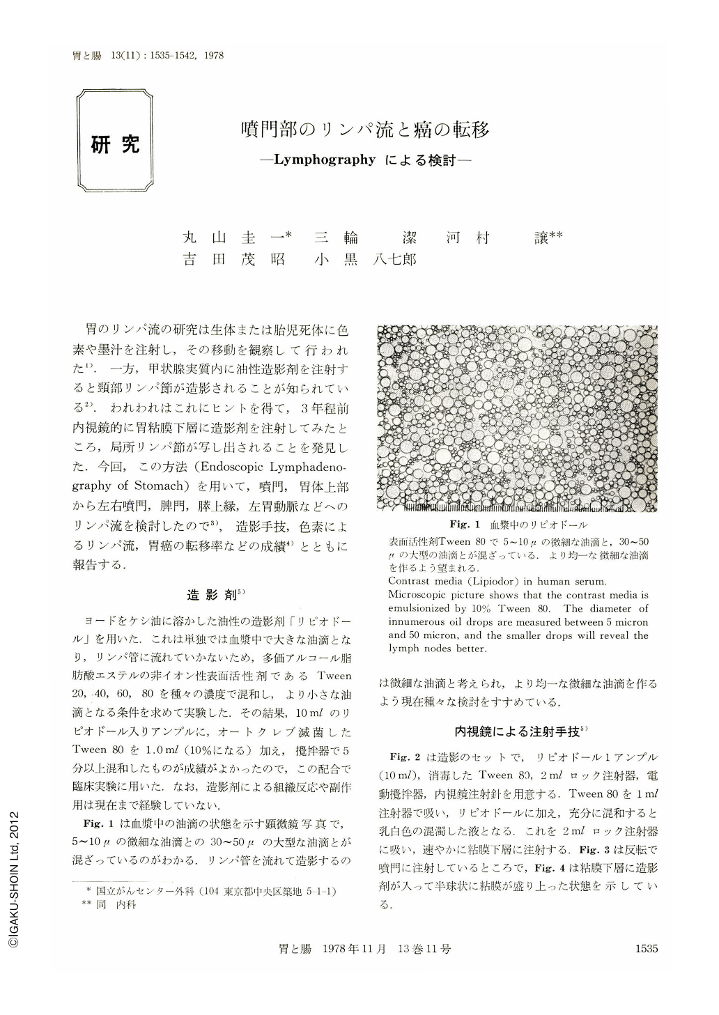
ヨードをケシ油に溶かした油性の造影剤「リピオドール」を用いた.これは単独では血漿中で大きな油滴となり,リンパ管に流れていかないため,多価アルコール脂肪酸エステルの非イオン性表面活性剤であるTween20,40,60,80を種々の濃度で混和し,より小さな油滴となる条件を求めて実験した.その結果,10mlのリピオドール入りアンプルに,オートクレブ滅菌したTween80を1.0ml(10%になる)加え,攪拌器で5分以上混和したものが成績がよかったので,この配合で臨床実験に用いた.なお,造影剤による組織反応や副作用は現在まで経験していない.
Taking advantage of the merit that confluent lymph nodes could be visualized by the endoscopic injection of oily contrast media into the submucosal layer, we observed lymphatic flow from the gastric cardia and upper part of the corpus. We also compared the results with the lymphatic flow as was visualized by dye injection and with the incidence of lymph node metastasis of gastric cancer.
Lymphography can be made by accurate endoscopic injection of 2~3 ml of Lipiodol emulsionized with Tween 20 into the submucosal layer of the upper body. Plain films of the abdomen were taken one and four days after injection and we conjectured the lymphatic flow by judging how lymph nodes were visualized through exposures of the resected material at operation and especially of the excised lymph nodes. We also observed the dye (Patento blue) circulating in the lymphatic vessels after it had been injected during operation into the subserosal side of the site of lymphography. The results of lymphography were also compared with the rate of metastasis of each lymph node in patients with gastric cancer localized in the C region.
Lymphography was done in 15 patients. The results of the above-mentioned three studies corresponded well each other. In conclusion, we have assumed that there be three lymphatic flow systems arising from the upper part of the stomach. They are: ―
A. Ascending flow reaching the mediastium along the esophageal wall.
B. Right-side flow from the lesser curvature through cardio-esophageal branch and left gastric artery to the celiac artery.
C. Left-side flow from the posterior wall through the greater curvature and the upper border of the pancreas to the retroperitoneum. Left-side flow is further subdivided into C-1: greater curvature channel from the greater curvature through short gastric arteries, splenic hilus and splenic artery to celiac artery; C-2: posterior gastric channel flowing from the posterior wall of the stomach along ramus esophagogastricus posterior ascendens joining the splenic artery system on the upper margin of the pancreas; C-3: diaphragmatic channel flowing from the fornix, left cardia along the left inferior phrenic artery (esophagocardiac branch) joing directly the para-aortic lymphatics. Our results are considered of great help in lymph node dissection of gastric cancer.
Copyright © 1978, Igaku-Shoin Ltd. All rights reserved.