Japanese
English
- 有料閲覧
- Abstract 文献概要
- 1ページ目 Look Inside
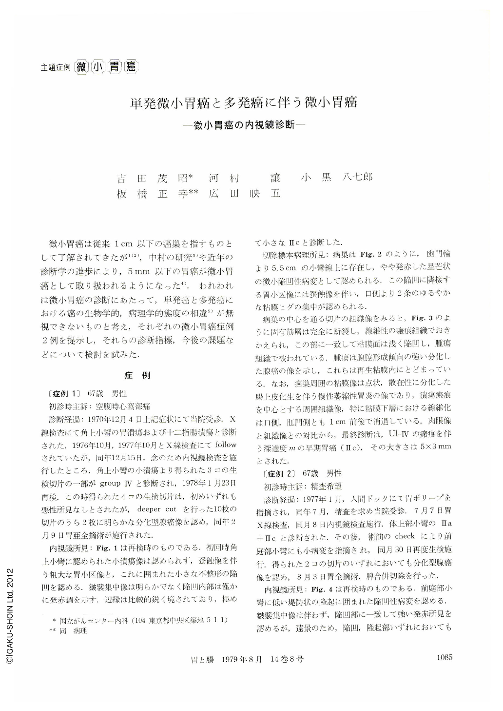
微小胃癌は従来1cm以下の癌巣を指すものとして了解されてきたが1)2),中村の研究3)や近年の診断学の進歩により,5mm以下の胃癌が微小胃癌として取り扱われるようになった4).われわれは微小胃癌の診断にあたって,単発癌と多発癌における癌の生物学的,病理学的態度の相違5)が無視できないものと考え,それぞれの微小胃癌症例2例を提示し,それらの診断指標,今後の課題などについて検討を試みた.
We demonstrated 2 cases of minute gastric cancer, which measured less than 5mm in diameter. The lesion of case 1 was solitary and that of case 2 was found as a component of multiple gastric carcinoma. Both were Ⅱc type of early gastric cancer whose invasion was limited in mucosal layer and the histological type was well differentiated adenocarcinoma.
The endoscopic appearances of these lesions were similar at first sight, and they were minute reddish depression surrounded with slight elevation. Close observation, however, revealed some differences between the two. In ease 1, a slight convergence of the mucosal folds and moth-eaten signs on the areagastricae which surrounded the depression indicated the presence of ulcerative change. Histological examination showed an ulcer scar grade Ⅳ (Murakami) in this lesion. On the other hand, case 2 looked like a minute form of Ⅱa+Ⅱc type early gastric cancer because of depression with annular elevation. And histology revealed no ulcerative change in this lesion.
In our previous study, 900 cases of early gastric cancer were examined on the multiplicity of lesions. In 823 of 900 cases, cancer was solitary and in 73 multiple. As shown in Fig. 7, in comparison with solitary cancer, the multiple gastric cancer was seen more often in male and the predominant histological type was well differentiated adenocarcinoma. And in most case of multiple gastric cancer, the incidence of histologically proved ulcerative change was low and local invasion was limited in mucosal layer. These differences between solitary and multiple early gastric cancer were statistically significant. And the similar comparative study carried on 63 cases of small gastric cancer (less than 10 mm in diameter) showed identical differences between solitary and multiple lesions. From these results, we assumed that the differences are same regardless of the size of lesions, even in minute cancer under 5mm in diameter.
The differences in the endoscopic view of the minute cancer cases reported here show those of the nature of solitary and multiple carcinoma.
For the endoscopical detection of solitary minute cancer, the retrospective studies of a minute cancer found unexpectedly in multiple cancer lesion alone seems to be insufficient. Therefore detailed investigation of the endoscopic appearances of solitary gastric cancer (>5 mm included), especially the cases whose presence are difficult to confirm, must be imperative.
At present, the most common endoscopical changes in solitary minute cancer are supposed to be: 1) indistinct shallow depression with red granular changes.
2) gradually converging folds toward discolored area.
3) coarse area-gastricae very similar to surrounding mucosa.
In future, detailed examination of parameters of malignancy must be carried on not only in gross change like the abrupt disappearance of folds but also in gastritis-like minute changes.

Copyright © 1979, Igaku-Shoin Ltd. All rights reserved.


