Japanese
English
- 有料閲覧
PPV購入案内
1,320円 (1,200円+税10%) こちらは、1531ページから1535ページの文献です。購入には医書.jp本会員登録が必要です。
会員登録完了後に改めて上記「購入サイトへ」を選択してください。
または「購入サイトへ」選択後に「購入ログイン」、「新規会員登録」の順に進んでください。
会員登録について詳しくはをご確認ください。
- Abstract 文献概要
- 1ページ目 Look Inside
- サイト内被引用 Cited by
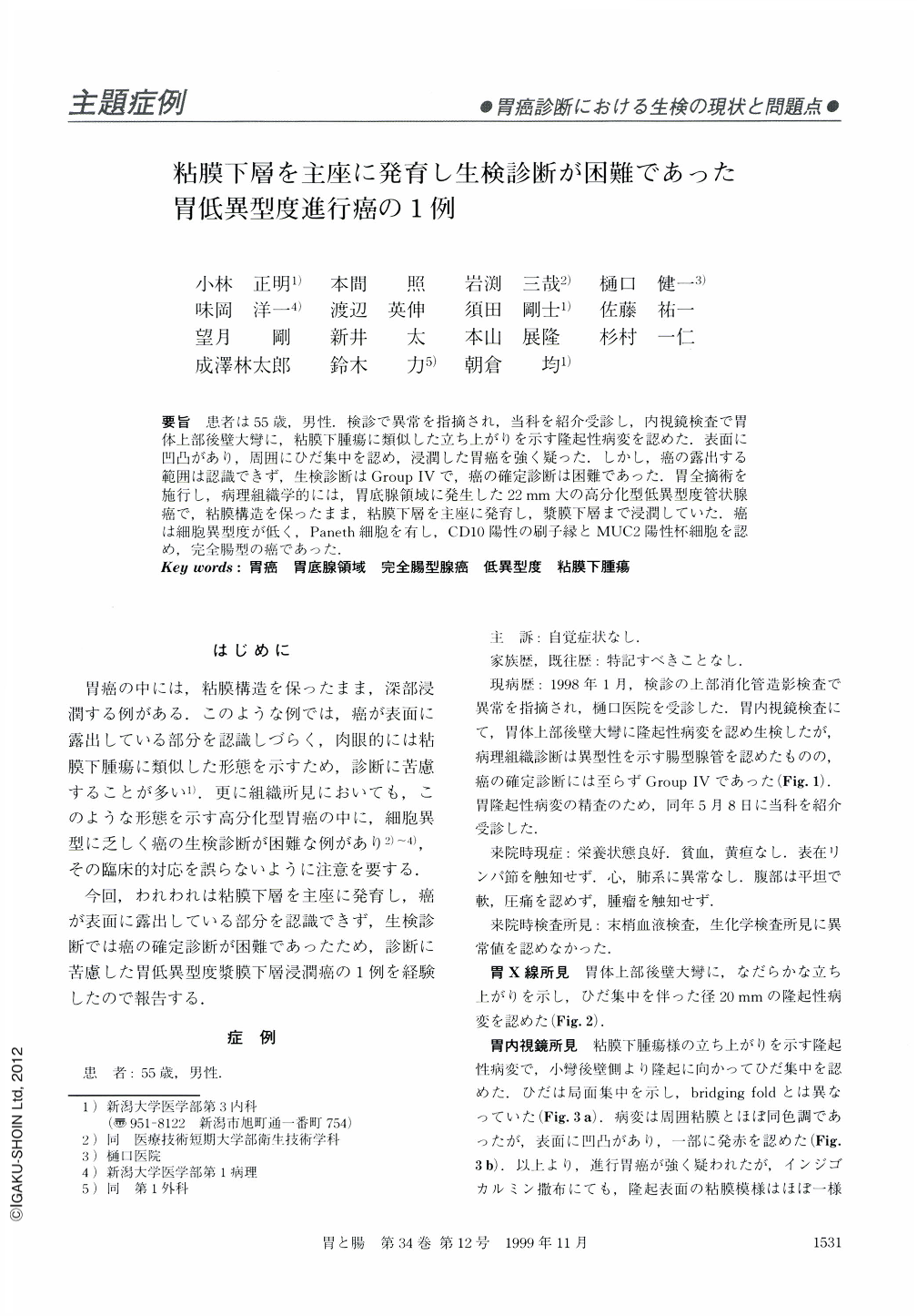
要旨 患者は55歳,男性.検診で異常を指摘され,当科を紹介受診し,内視鏡検査で胃体上部後壁大彎に,粘膜下腫瘍に類似した立ち上がりを示す隆起性病変を認めた.表面に凹凸があり,周囲にひだ集中を認め,浸潤した胃癌を強く疑った.しかし,癌の露出する範囲は認識できず,生検診断はGroup Ⅳで,癌の確定診断は困難であった.胃全摘術を施行し,病理組織学的には,胃底腺領域に発生した22mm大の高分化型低異型度管状腺癌で,粘膜構造を保ったまま,粘膜下層を主座に発育し,漿膜下層まで浸潤していた.癌は細胞異型度が低く,Paneth細胞を有し,CD10陽性の刷子縁とMUC2陽性杯細胞を認め,完全腸型の癌であった.
We report a case of gastric carcinoma with submucosal growth. A 55-year-old man underwent upper gastroendoscopic examination, which detected a 22 mm sized protruding tumor at the posterior wall of the upper body. The tumor was covered with normal-looking mucosa, but showed nodular surface and fold convergence. Histological findings revealed such low-grade atypia that biopsy examination resulted in a diagnosis of Group Ⅳ, in which the diagnosis for carcinoma is inconclusive. The patient underwent surgical resection. Pathological diagnosis was well differentiated adenocarcinoma of the complete intestinal phenotype, invading to the subserosa. The carcinoma arose from the fundic gland mucosa.
Copyright © 1999, Igaku-Shoin Ltd. All rights reserved.