Japanese
English
- 有料閲覧
- Abstract 文献概要
- 1ページ目 Look Inside
- サイト内被引用 Cited by
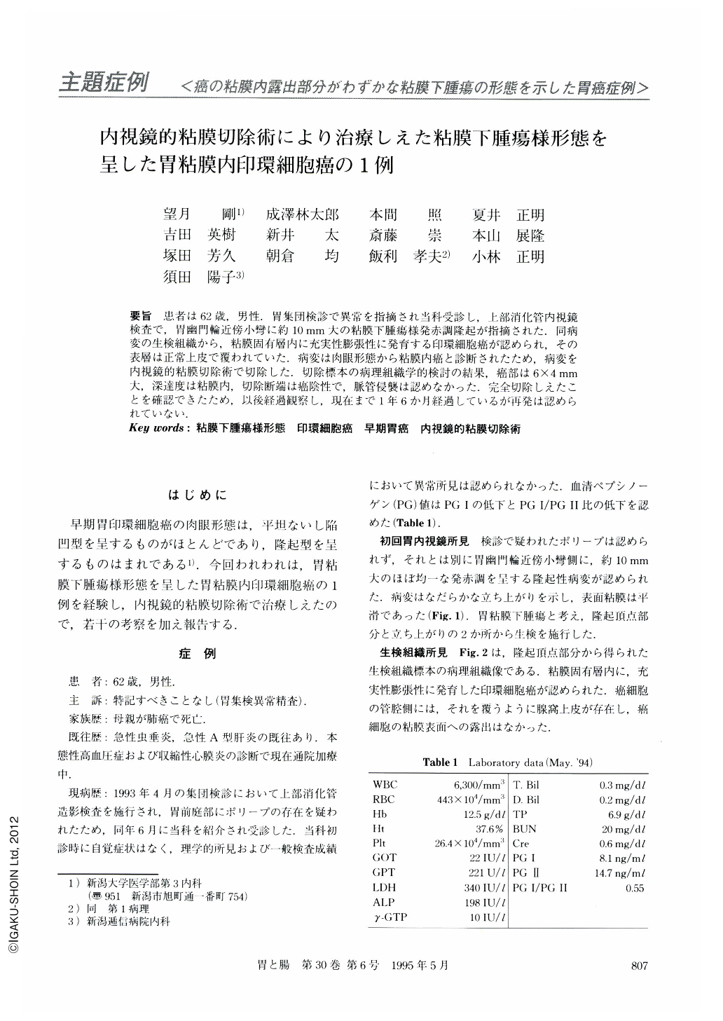
要旨 患者は62歳,男性.胃集団検診で異常を指摘され当科受診し,上部消化管内視鏡検査で,胃幽門輪近傍小彎に約10mm大の粘膜下腫瘍様発赤調隆起が指摘された.同病変の生検組織から,粘膜固有層内に充実性膨張性に発育する印環細胞癌が認められ,その表層は正常上皮で覆われていた.病変は肉眼形態から粘膜内癌と診断されたため,病変を内視鏡的粘膜切除術で切除した.切除標本の病理組織学的検討の結果,癌部は6×4mm大,深達度は粘膜内,切除断端は癌陰性で,脈管侵襲は認めなかった.完全切除しえたことを確認できたため,以後経過観察し,現在まで1年6か月経過しているが再発は認められていない.
A 62-year-old man visited our hospital for further examination of an abnormality of the stomach found at a mass gastric survey. On endoscopic examination at our hospital, a submucosal tumor-like lesion, about 10 mm in diameter, was found on the lesser curvature near the pyloric ring. Signet-ring cell carcinoma was revealed in the biopsy specimen taken from top of the lesion. These cancer cells were covered by epithelia, grew medullarily and expansively, and existed in the middle and deep layers of the mucosa. Endoscopic mucosal resection was carried out successfully. Histological examination revealed a signet-ring cell carcinoma limited to the mucosa, measuring 6×4 mm in size, and the lesion was completely resected without lymphatic or vascular permeation.

Copyright © 1995, Igaku-Shoin Ltd. All rights reserved.


