Japanese
English
- 有料閲覧
- Abstract 文献概要
- 1ページ目 Look Inside
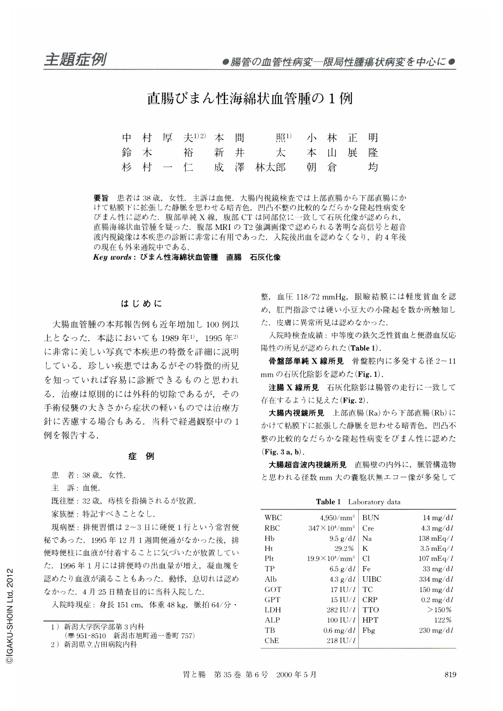
要旨 患者は38歳,女性.主訴は血便.大腸内視鏡検査では上部直腸から下部直腸にかけて粘膜下に拡張した静脈を思わせる暗青色,凹凸不整の比較的なだらかな隆起性病変をびまん性に認めた.腹部単純X線,腹部CTは同部位に一致して石灰化像が認められ,直腸海綿状血管腫を疑った.腹部MRIのT2強調画像で認められる著明な高信号と超音波内視鏡像は本疾患の診断に非常に有用であった.入院後出血を認めなくなり,約4年後の現在も外来通院中である.
A 38-year-old woman with a complaint of constipation and occasional melena was referred to our hospital. Colonoscopy revealed bluish meandering SMT-like lesions resembling rectal varices in the distal rectum, circumferentially. Plain abdominal x-ray examination showed multiple small calcifications in the pelvic cavity. Endscopic ultrasonography was useful for diagnosis of microcystic lesions and echogenic spots, indicating dilated vessels and small calcifications. After prescription of laxtive, the patient's hematochezia has improved and no bloody stool has been seen.

Copyright © 2000, Igaku-Shoin Ltd. All rights reserved.


