Japanese
English
- 有料閲覧
PPV購入案内
1,320円 (1,200円+税10%) こちらは、989ページから999ページの文献です。購入には医書.jp本会員登録が必要です。
会員登録完了後に改めて上記「購入サイトへ」を選択してください。
または「購入サイトへ」選択後に「購入ログイン」、「新規会員登録」の順に進んでください。
会員登録について詳しくはをご確認ください。
- Abstract 文献概要
- 1ページ目 Look Inside
- 参考文献 Reference
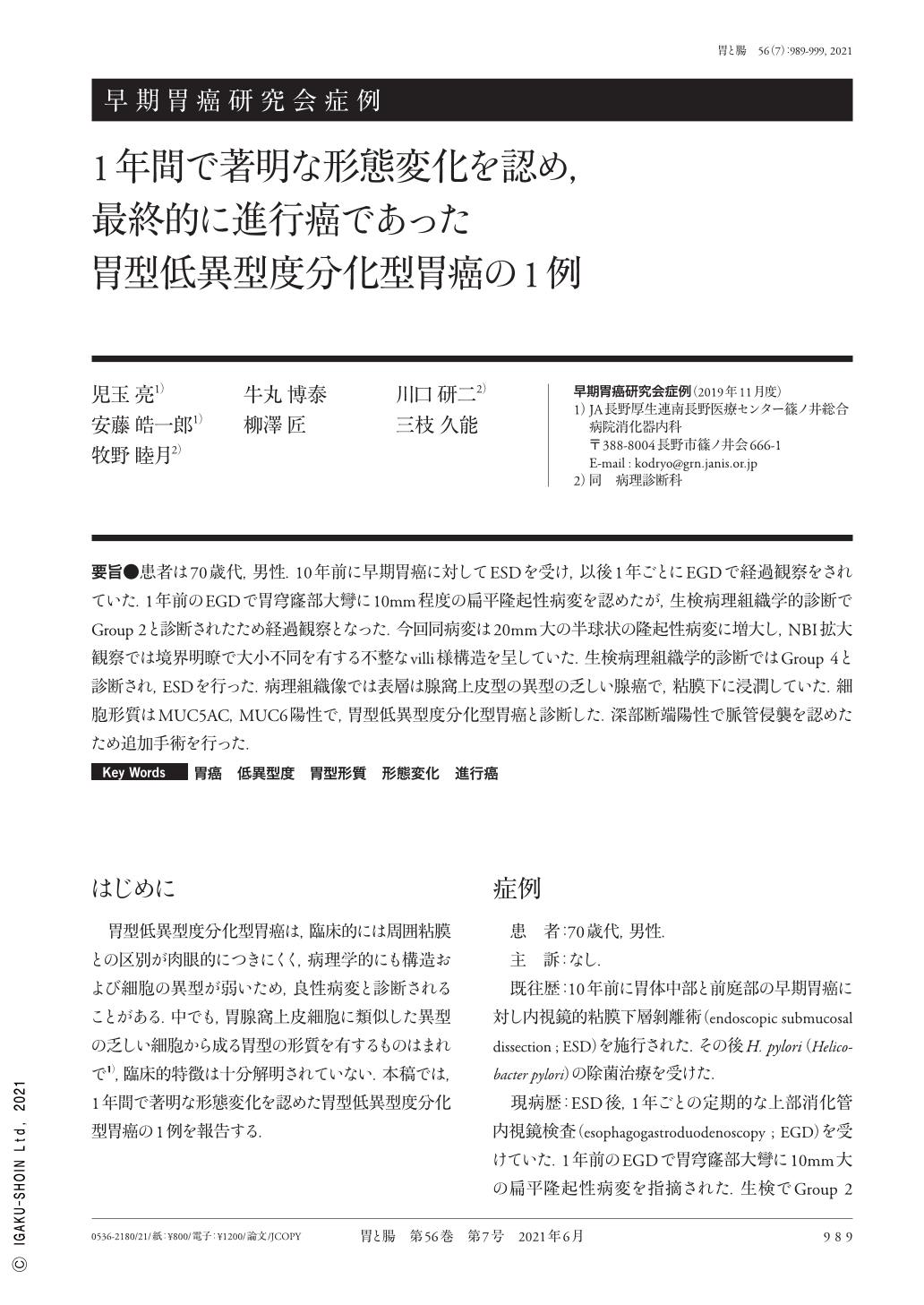
要旨●患者は70歳代,男性.10年前に早期胃癌に対してESDを受け,以後1年ごとにEGDで経過観察をされていた.1年前のEGDで胃穹窿部大彎に10mm程度の扁平隆起性病変を認めたが,生検病理組織学的診断でGroup 2と診断されたため経過観察となった.今回同病変は20mm大の半球状の隆起性病変に増大し,NBI拡大観察では境界明瞭で大小不同を有する不整なvilli様構造を呈していた.生検病理組織学的診断ではGroup 4と診断され,ESDを行った.病理組織像では表層は腺窩上皮型の異型の乏しい腺癌で,粘膜下に浸潤していた.細胞形質はMUC5AC,MUC6陽性で,胃型低異型度分化型胃癌と診断した.深部断端陽性で脈管侵襲を認めたため追加手術を行った.
A 70-year-old man underwent endoscopic submucosal dissection for early gastric cancer 10 years ago, following which he received esophagogastroduodenoscopy every year. During follow-up, a flat elevated lesion measuring 10mm in size was detected in the greater curvature at the fornix of the stomach. Biopsy specimens were diagnosed as group 2. After 1 year, the lesion progressed to a hemispherical elevated lesion measuring 20mm in size. Narrow-band imaging with magnification revealed an irregular mucosal surface pattern of villi-like structure. Based on the biopsy specimens, an extremely well-differentiated adenocarcinoma was suspected. We performed endoscopic submucosal dissection. A pathological examination revealed that the superficial layer of the lesion was very mild atypia of foveolar-type adenocarcinoma, and the tumor invaded to the submucosal layer. Furthermore, the tumor cells were positive for MUC5AC and MUC6 ; therefore, we diagnosed the lesion as a gastric-type, low-grade, well-differentiated adenocarcinoma of the stomach. The vertical margin of the resected specimens was positive with the involvement of vessels, and the patient was subsequently subjected to total gastrectomy.
Copyright © 2021, Igaku-Shoin Ltd. All rights reserved.