Japanese
English
- 有料閲覧
- Abstract 文献概要
- 1ページ目 Look Inside
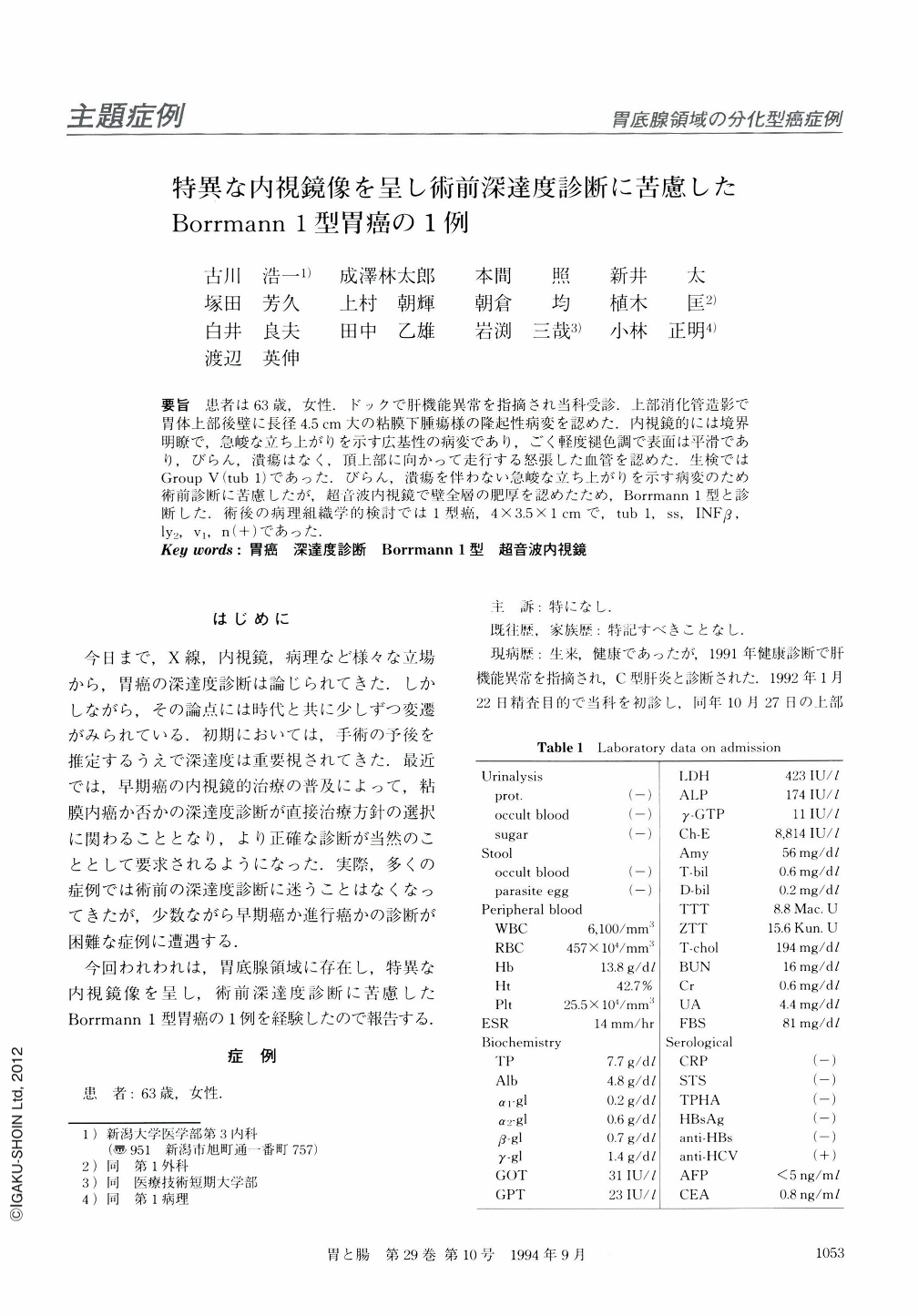
要旨 患者は63歳,女性.ドックで肝機能異常を指摘され当科受診.上部消化管造影で胃体上部後壁に長径4.5cm大の粘膜下腫瘍様の隆起性病変を認めた.内視鏡的には境界明瞭で,急峻な立ち上がりを示す広基性の病変であり,ごく軽度褪色調で表面は平滑であり,びらん,潰瘍はなく,頂上部に向かって走行する怒張した血管を認めた.生検ではGroup Ⅴ(tub1)であった.びらん,潰瘍を伴わない急峻な立ち上がりを示す病変のため術前診断に苦慮したが,超音波内視鏡で壁全層の肥厚を認めたため,Borrmann1型と診断した.術後の病理組織学的検討では1型癌,4×3.5×1cmで,tubl,ss,INFβ,ly2,V1,n(+)であった.
We encountered a 63-year-old woman who, while undergoing a thorough health screen, was found to have abnormalities in liver function. Roentgenologic examination of the upper gastrointestinal tract on admis sion revealed an exphytic neoplasm of 4.5 cm in diameter located on the posterior wall of the gastric body. Endoscopically, the tumor was a fungating one with broad base and well-defined boundaries. It was faintly brownish in color with smooth surfaces and without erosion or ulceration; a tortuous blood vessel running towards the apex of the tumor was observed. Biopsy yielded findings that were compatible with a Group V (tub1) carcinoma. The unique morphologic features perplexed us when making a preoperative diagnosis of the depth of invasion. Ultrasound endoscopy disclosed thickening of entire layers of the affected stomach wall, thus leading to a conclusive diagnosis of Borrmann type l carcinoma. Histologic examination of involved tissue resected at operation allowed us to determine the depth of invasion as ss.

Copyright © 1994, Igaku-Shoin Ltd. All rights reserved.


