Japanese
English
- 有料閲覧
PPV購入案内
1,320円 (1,200円+税10%) こちらは、1513ページから1520ページの文献です。購入には医書.jp本会員登録が必要です。
会員登録完了後に改めて上記「購入サイトへ」を選択してください。
または「購入サイトへ」選択後に「購入ログイン」、「新規会員登録」の順に進んでください。
会員登録について詳しくはをご確認ください。
- Abstract 文献概要
- 1ページ目 Look Inside
- サイト内被引用 Cited by
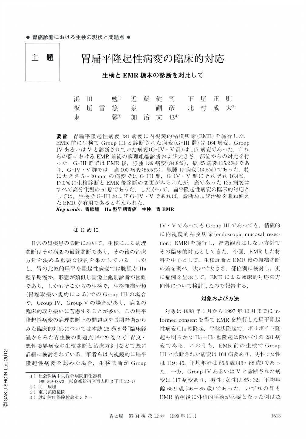
要旨 胃扁平隆起性病変281病変に内視鏡的粘膜切除(EMR)を施行した.EMR前に生検でGroupⅢと診断された病変(G-Ⅲ群)は164病変,GroupⅣあるいはⅤと診断されていた病変(G-Ⅳ・Ⅴ群)は117病変であった.これらの群におけるEMR前後の病理組織診断および大きさ,部位からの対比を行った.G-Ⅲ群ではEMR後,腺腫139病変(84.8%),癌25病変(152%)であり,G-Ⅳ・Ⅴ群では,癌100病変(85.5%),腺腫17病変(14.5%)であった.特に大きさ5~20mmの病変ではG-Ⅲ群,G-Ⅳ・Ⅴ群にそれぞれ16.4%,17.O%に生検診断とEMR後診断の変更がみられたが,癌であった125病変はすべて高分化型のm癌であった.したがって,扁平隆起性病変の臨床的対応としては,生検でG-ⅢおよびG-Ⅳ・Ⅴであれば,診断および治療を兼ね備えたEMRが有用であると考えられた.
Usually, we confirm the diagnosis of a gastric mucosal lesion by endoscopy and biopsy specimen. During the period from January, 1988 to December, 1997, 164 flat elevated lesions diagnosed as Group Ⅲ and 117 lesions diagnosed as Group Ⅳ ・ Ⅴ by the biopsy specimen were treated by endoscopical mucosal resection (EMR) in our department. We discussed how to manage flat elevated lesions clinically.
In the Group Ⅲ lesions, 139 lesions (84.8%) were diagnosed as gastric adenoma and 25 lesions (15.2%) were diagnosed as gastric cancer by histological examination of the resected specimens. On the other hand, in the Group Ⅳ ・ Ⅴ lesions, 100 lesions (85.5%) were diagnosed as cancer and 17 lesions (14.5%) were diagnosed as adenoma. The rate of difference between the histological diagnoses based on biopsy specimens and those based on resected specimens by EMR had no relation to the size and the location of the lesions. After histological examination of specimens obtained by EMR, the origind diagnoses were changed in about 15% of the cases. Those results suggested that the diagnosis of flat elevated lesions were difficult not only macroscopically but also histologically. Considering this, from a clinical viewpoint, we concluded that, EMR should be performed for lesions which are endoscopically diagnosed as flat elevated lesions, irrespectively of the whether the biopsies indicate they belong to Group Ⅲ, Ⅳ or Ⅴ.
Copyright © 1999, Igaku-Shoin Ltd. All rights reserved.