Japanese
English
- 有料閲覧
PPV購入案内
1,320円 (1,200円+税10%) こちらは、47ページから56ページの文献です。購入には医書.jp本会員登録が必要です。
会員登録完了後に改めて上記「購入サイトへ」を選択してください。
または「購入サイトへ」選択後に「購入ログイン」、「新規会員登録」の順に進んでください。
会員登録について詳しくはをご確認ください。
- Abstract 文献概要
- 1ページ目 Look Inside
- サイト内被引用 Cited by
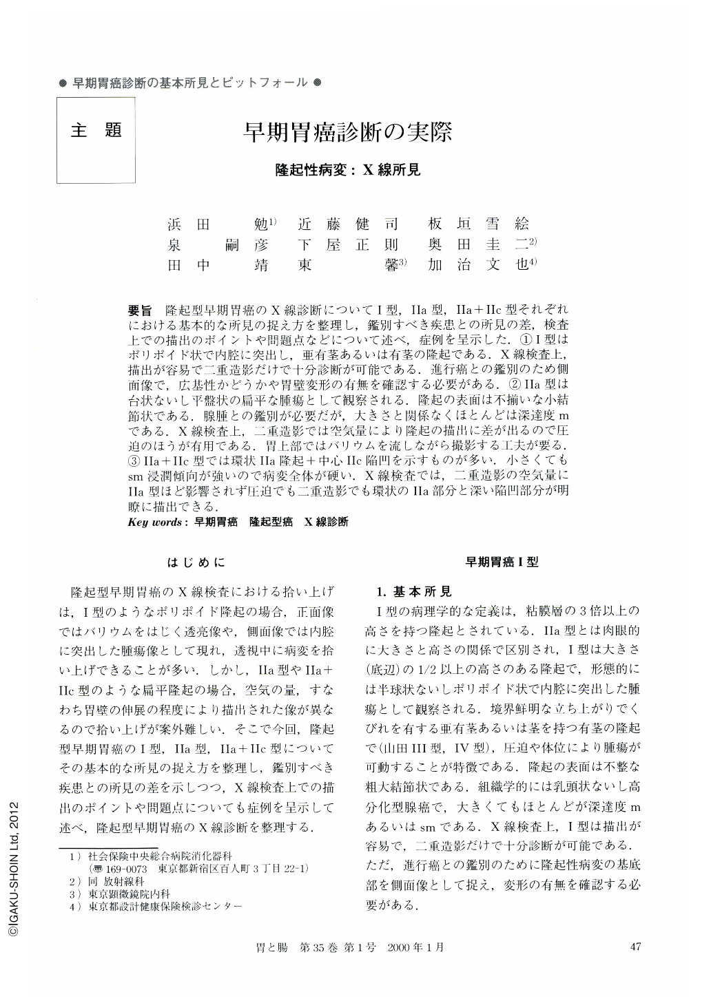
要旨 隆起型早期胃癌のX線診断についてI型,Ⅱa型,Ⅱa+Ⅱc型それぞれにおける基本的な所見の捉え方を整理し,鑑別すべき疾患との所見の差,検査上での描出のポイントや問題点などについて述べ,症例を呈示した.①1型はポリポイド状で内腔に突出し,亜有茎あるいは有茎の隆起である.X線検査上,描出が容易で二重造影だけで十分診断が可能である.進行癌との鑑別のため側面像で,広基性かどうかや胃壁変形の有無を確認する必要がある.②Ⅱa型は台状ないし平盤状の扁平な腫瘍として観察される.隆起の表面は不揃いな小結節状である.腺腫との鑑別が必要だが,大きさと関係なくほとんどは深達度mである.X線検査上,二重造影では空気量により隆起の描出に差が出るので圧迫のほうが有用である.胃上部ではバリウムを流しながら撮影する工夫が要る.③Ⅱa+Ⅱc型では環状Ⅱa隆起+中心Ⅱc陥凹を示すものが多い.小さくてもsm浸潤傾向が強いので病変全体が硬い.X線検査では,二重造影の空気量にⅡa型ほど影響されず圧迫でも二重造影でも環状のⅡa部分と深い陥凹部分が明瞭に描出できる.
The macroscopic classification of polypoid early cancer includes three types; namely type I (protuded type), type Ⅱa (superficial, elevated type) and type Ⅱa+Ⅱc. In this paper, the basic radiographic findings were described in order to distinguish the three types from other polypoid lesions.
(1) Type I cancers are characterized by a pedunculated or subpedunculated contour with marked protrusion into the gastric lumen, so it is easy to diagnose them either by a double contrast or a compression image. But, a polypoid tumor with a wide-base and with rigidity of the gastric wall on the radiographic lateral view is suggestive of advanced cancer.
(2) Type Ⅱa is recognized by its having a flat elevation with a sessile profile form and in most cases of this type there is cancerous invasion limited to the mucosa with no relation to size. The granular surface pattern is characteristic of this type, but, it's hard to distinguish it from adenoma. The compression views on radiography usually give better pictures than double contrast views. In case of Ⅱa situated in the upper part of the stomach radiolucent shadow should be taken while observing the flow of the barium during the examination.
(3) Most type Ⅱa+Ⅱc cancers are characterized by a central Ⅱc-like depression within the Type Ⅱa. Most cancers of this type easily invade to the submucosa even though they are less than 10 mm in size. Cancers with submucosal invasion become hard, so both the compression and the double contrast examination are reliable methods for delineating all their aspects.
Copyright © 2000, Igaku-Shoin Ltd. All rights reserved.