Japanese
English
- 有料閲覧
- Abstract 文献概要
- 1ページ目 Look Inside
- サイト内被引用 Cited by
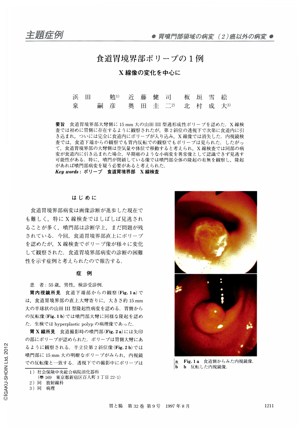
要旨 食道胃境界部大彎側に15mm大の山田III型過形成性ポリープを認めた.X線検査では初めに胃側に存在するように観察されたが,第2斜位の透視下で次第に食道内に引き込まれ,ついには完全に食道内にポリープが入り込み,X線像では消失した.内視鏡検査では,食道下端からの観察でも胃内反転での観察でもポリープは見られた.したがって,食道胃境界部の大彎側は空気量や体位で移動すると考えられ,X線検査では同部の病変が食道内に引き込まれた場合,早期癌のような小病変を異常像として認識できず見逃す可能性がある.特に,噴門が閉鎖している像では噴門部全体の隆起の有無を観察し,隆起があれば噴門部病変を疑う必要があると考えられた.
During a regular check-up, a gastric polyp at the esophago-gastric junction of a 55-year-old man was discovered by X-ray. The polyp, 15 mm in size, was recognized as a radiolucent shadow at the semi-standing leftanterior oblique position. But the shadow disappered at the same position, when the cardia was completely closed during the examination. It suggested that the polyp was drawn into the esophgus side from the gastric side because of the movement of the esophagogastric junction.
At endoscopic examination, a hemispheric polyp without stalks (Yamada's classification, type III) was observed just over the esophago‐gastric junction and the biopsy specimen was identified as a hyperplastic benign polyp.
It is thought that the esophago-gastric junction moves occasionaly from the gastric side to the esophageal side. On account of this, when we read the X-ray films, we should be aware that a small polypoid lesion at the esophago-gastric junction might exist even though there is no radiolucent shadow on X-ray films.

Copyright © 1997, Igaku-Shoin Ltd. All rights reserved.


