Japanese
English
- 有料閲覧
PPV購入案内
1,320円 (1,200円+税10%) こちらは、579ページから586ページの文献です。購入には医書.jp本会員登録が必要です。
会員登録完了後に改めて上記「購入サイトへ」を選択してください。
または「購入サイトへ」選択後に「購入ログイン」、「新規会員登録」の順に進んでください。
会員登録について詳しくはをご確認ください。
- Abstract 文献概要
- 1ページ目 Look Inside
- 参考文献 Reference
- サイト内被引用 Cited by
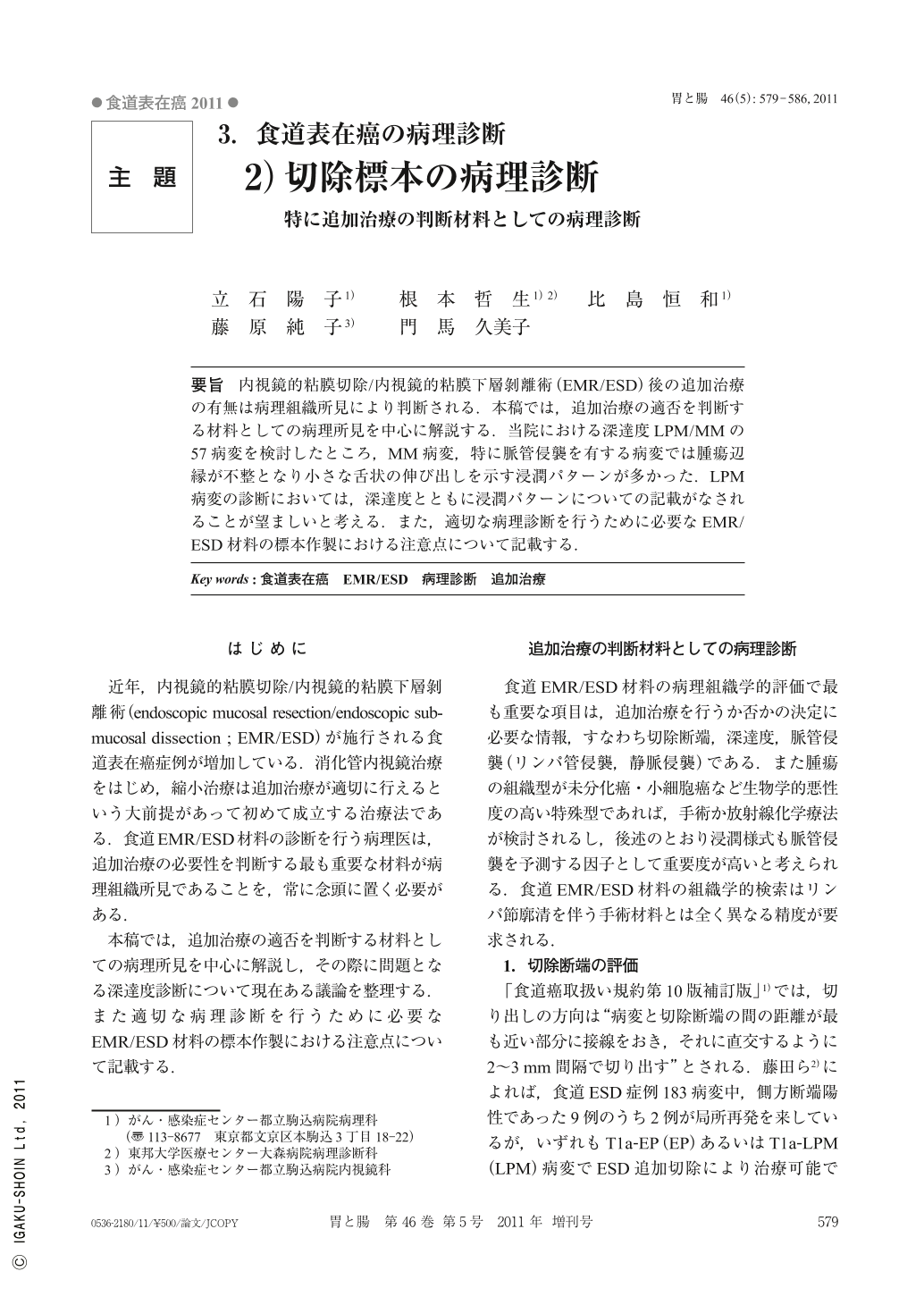
要旨 内視鏡的粘膜切除/内視鏡的粘膜下層剝離術(EMR/ESD)後の追加治療の有無は病理組織所見により判断される.本稿では,追加治療の適否を判断する材料としての病理所見を中心に解説する.当院における深達度LPM/MMの57病変を検討したところ,MM病変,特に脈管侵襲を有する病変では腫瘍辺縁が不整となり小さな舌状の伸び出しを示す浸潤パターンが多かった.LPM病変の診断においては,深達度とともに浸潤パターンについての記載がなされることが望ましいと考える.また,適切な病理診断を行うために必要なEMR/ESD材料の標本作製における注意点について記載する.
The necessity of additional treatment after EMR/ESD(endoscopic mucosal resection/endoscopic submucosal dissection)is evaluated by histopathological findings. Here, we mainly discuss histopathological findings as a means of evaluating the necessity of additional treatment. In 57 lesions with LPM/MM invasion, all cases of MM lesions with invasion to the vessels, showed an infiltration pattern in which the tumor invasive front was irregular and extended into interstitium like small buds. For diagnosis of LPM lesions, it is considered desirable to describe the infiltration pattern as well as the depth of invasion. In addition, points requiring caution in preparing specimens of EMR/ESD materials, which are necessary for appropriate pathological diagnoses, will be presented.
Copyright © 2011, Igaku-Shoin Ltd. All rights reserved.