Japanese
English
- 有料閲覧
- Abstract 文献概要
- 1ページ目 Look Inside
- 参考文献 Reference
- サイト内被引用 Cited by
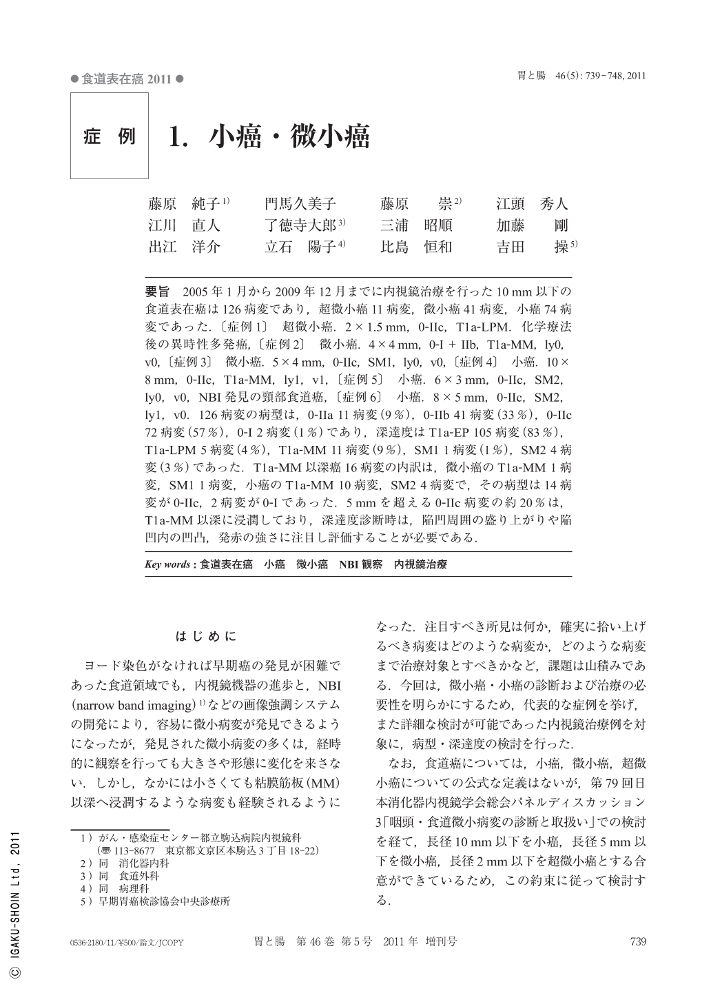
要旨 2005年1月から2009年12月までに内視鏡治療を行った10mm以下の食道表在癌は126病変であり,超微小癌11病変,微小癌41病変,小癌74病変であった.〔症例1〕 超微小癌.2×1.5mm,0-IIc,T1a-LPM.化学療法後の異時性多発癌,〔症例2〕 微小癌.4×4mm,0-I+IIb,T1a-MM,ly0,v0,〔症例3〕 微小癌.5×4mm,0-IIc,SM1,ly0,v0,〔症例4〕 小癌.10×8mm,0-IIc,T1a-MM,ly1,v1,〔症例5〕 小癌.6×3mm,0-IIc,SM2,ly0,v0,NBI発見の頸部食道癌,〔症例6〕 小癌.8×5mm,0-IIc,SM2,ly1,v0.126病変の病型は,0-IIa 11病変(9%),0-IIb 41病変(33%),0-IIc 72病変(57%),0-I 2病変(1%)であり,深達度はT1a-EP 105病変(83%),T1a-LPM 5病変(4%),T1a-MM 11病変(9%),SM1 1病変(1%),SM2 4病変(3%)であった.T1a-MM以深癌16病変の内訳は,微小癌のT1a-MM 1病変,SM1 1病変,小癌のT1a-MM 10病変,SM2 4病変で,その病型は14病変が0-IIc,2病変が0-Iであった.5mmを超える0-IIc病変の約20%は,T1a-MM以深に浸潤しており,深達度診断時は,陥凹周囲の盛り上がりや陥凹内の凹凸,発赤の強さに注目し評価することが必要である.
Total of 126 small superficial cancer lesions of the esophagus less than 10mm in size were noted among 476 esophageal cancer lesions(26.4%)underwent endoscopic treatment(EMR or ESD)from January 2005 to December 2009. Ultra minute cancers less than 2mm in size occupied 9% of all small lesions, minute cancers(2<x≦5mm)32% and small cancers(5<x≦10mm)59%. Type 0-IIa lesions occupied 9% of 126 lesions, type 0-IIb 33%, type 0-IIc 57% and type 0-I 1%. T1a-EP occupied 83% of 126 small cancer lesions, T1a-LPM 4%, T1a- MM 9%, T1b-SM1 1% and T1b-SM2 3%. In cases with ultra minute cancer lesions, cancer invasion was remained within T1a-LPM. While, minute cancer lesions occupied 12%(T1a-MM 6% and T1b-SM1 6%)of all 16 lesions with invasion of T1a-MM or more and small cancer lesion 88%(T1a-MM 63% and T1b-SM2 25%). At the same time, type 0-IIc lesions occupied 88% and type 0-I 12% of 16 cancer lesions with deeper invasion. In cases with type 0-IIc lesions more than 5mm in size, deeper invasion into the muscularis mucosae or more was noted in 20% of all cases. It is recommended in endoscopic evaluation of them that endoscopic findings suggesting deeper invasion such as slight elevation close to the margin, granular irregularities in the depression and partial and strong redness should be found out.

Copyright © 2011, Igaku-Shoin Ltd. All rights reserved.


