Japanese
English
- 有料閲覧
PPV購入案内
1,320円 (1,200円+税10%) こちらは、1131ページから1140ページの文献です。購入には医書.jp本会員登録が必要です。
会員登録完了後に改めて上記「購入サイトへ」を選択してください。
または「購入サイトへ」選択後に「購入ログイン」、「新規会員登録」の順に進んでください。
会員登録について詳しくはをご確認ください。
- Abstract 文献概要
- 1ページ目 Look Inside
- 参考文献 Reference
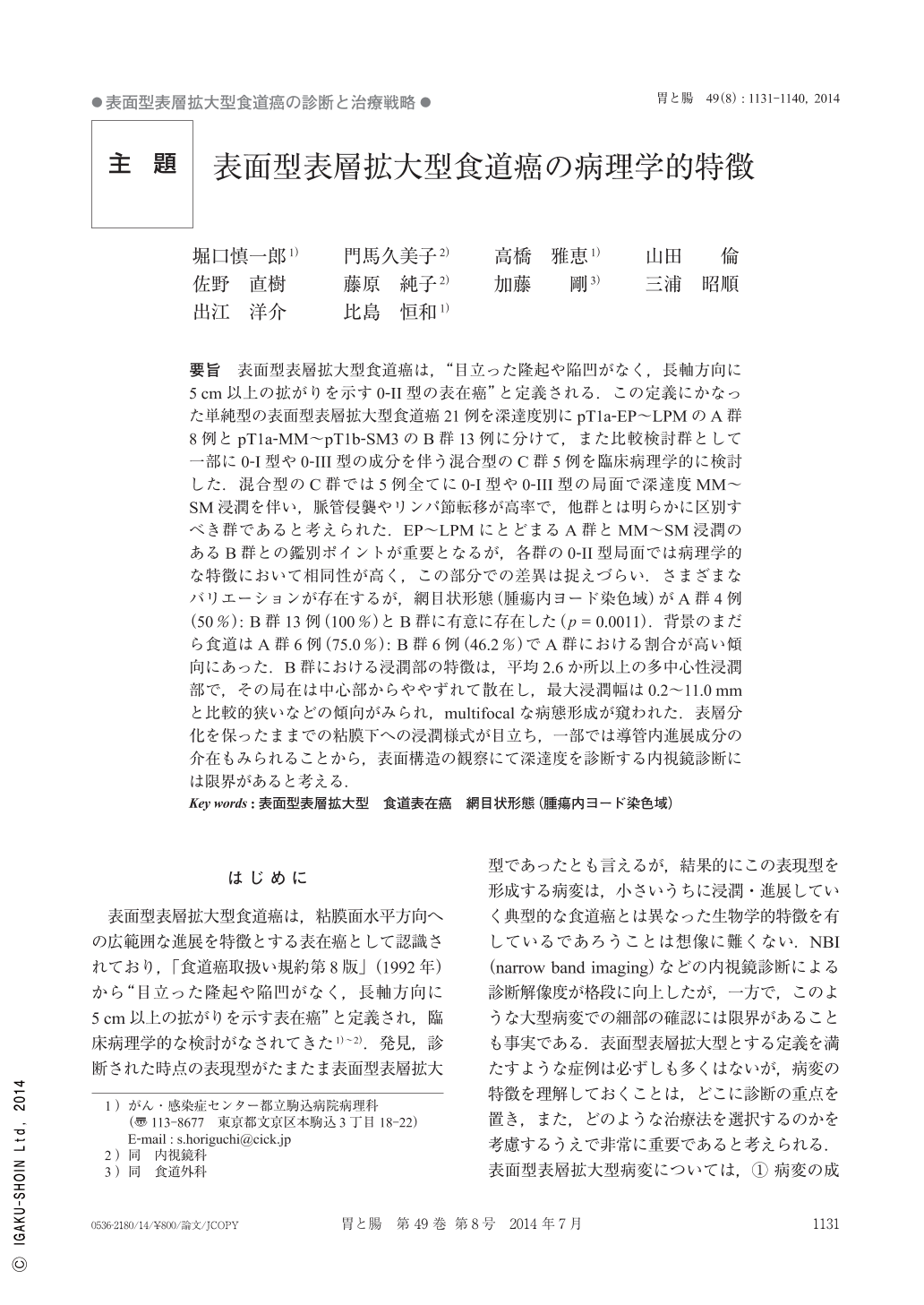
要旨 表面型表層拡大型食道癌は,“目立った隆起や陥凹がなく,長軸方向に5cm以上の拡がりを示す0-II型の表在癌”と定義される.この定義にかなった単純型の表面型表層拡大型食道癌21例を深達度別にpT1a-EP~LPMのA群8例とpT1a-MM~pT1b-SM3のB群13例に分けて,また比較検討群として一部に0-I型や0-III型の成分を伴う混合型のC群5例を臨床病理学的に検討した.混合型のC群では5例全てに0-I型や0-III型の局面で深達度MM~SM浸潤を伴い,脈管侵襲やリンパ節転移が高率で,他群とは明らかに区別すべき群であると考えられた.EP~LPMにとどまるA群とMM~SM浸潤のあるB群との鑑別ポイントが重要となるが,各群の0-II型局面では病理学的な特徴において相同性が高く,この部分での差異は捉えづらい.さまざまなバリエーションが存在するが,網目状形態(腫瘍内ヨード染色域)がA群4例(50%):B群13例(100%)とB群に有意に存在した(p=0.0011).背景のまだら食道はA群6例(75.0%):B群6例(46.2%)でA群における割合が高い傾向にあった.B群における浸潤部の特徴は,平均2.6か所以上の多中心性浸潤部で,その局在は中心部からややずれて散在し,最大浸潤幅は0.2~11.0mmと比較的狭いなどの傾向がみられ,multifocalな病態形成が窺われた.表層分化を保ったままでの粘膜下への浸潤様式が目立ち,一部では導管内進展成分の介在もみられることから,表面構造の観察にて深達度を診断する内視鏡診断には限界があると考える.
Superficial spreading-type esophageal carcinoma is defined as“0-II type superficial carcinomas that spread ≧5cm in the long axis of the esophagus without any prominent elevations or depressions.”On the basis of this definition, 21 patients with simple superficial-type spreading carcinoma of the esophagus were divided according to the depth of invasion into two groups : group A comprising 8 patients with pT1a-EP/LPM invasion and group B comprising 13 patients with pT1a-MM/pT1b-SM invasion. These 2 groups were clinicopathologically compared with a control group, group C, comprising 5 patients with combined type, including some patients with 0-III type and some others with 0-I type carcinomas. All group C patients had a high rate of lymph node metastasis and vascular invasion associated with MM/SM invasion at the 0-I and 0-III-type sites, which clearly distinguished them from the group A and B patients. A key point in distinguishing group A with EP/LPM invasion and group B with MM/SM invasion was that there was a strong homology in the pathological characteristics of both groups in terms of 0-II type aspects which were difficult to differentiate. Although there were variations within the groups, mesh-like forms(iodine-stained areas in the tumor)were prominent in 4 group A patients(50%)and 3 group B patients(100%)(p=0.0011). The esophagus was speckled in appearance in 5 group A patients(75.0%)and 6 group B patients(46.2%), and there was a high ratio in group A. Invasion in group B was characteristically multicentric, occurring at a mean of 2.6 sites, and the dissemination from the centers of lesions was relatively narrow with a maximum invasion diameter of 0.2~11mm, which was suggestive of a multifocal origin. Moreover, submucosal invasion was observed while differentiation remained superficial with some advanced mixed intraductal components. Our results suggest that the pathological characteristics of Group B type lesions should be carefully analyzed because of the possible factors complicating the endoscopic diagnosis of invasion depth according the surface appearance.
Copyright © 2014, Igaku-Shoin Ltd. All rights reserved.