Japanese
English
- 有料閲覧
- Abstract 文献概要
- 1ページ目 Look Inside
- 参考文献 Reference
- サイト内被引用 Cited by
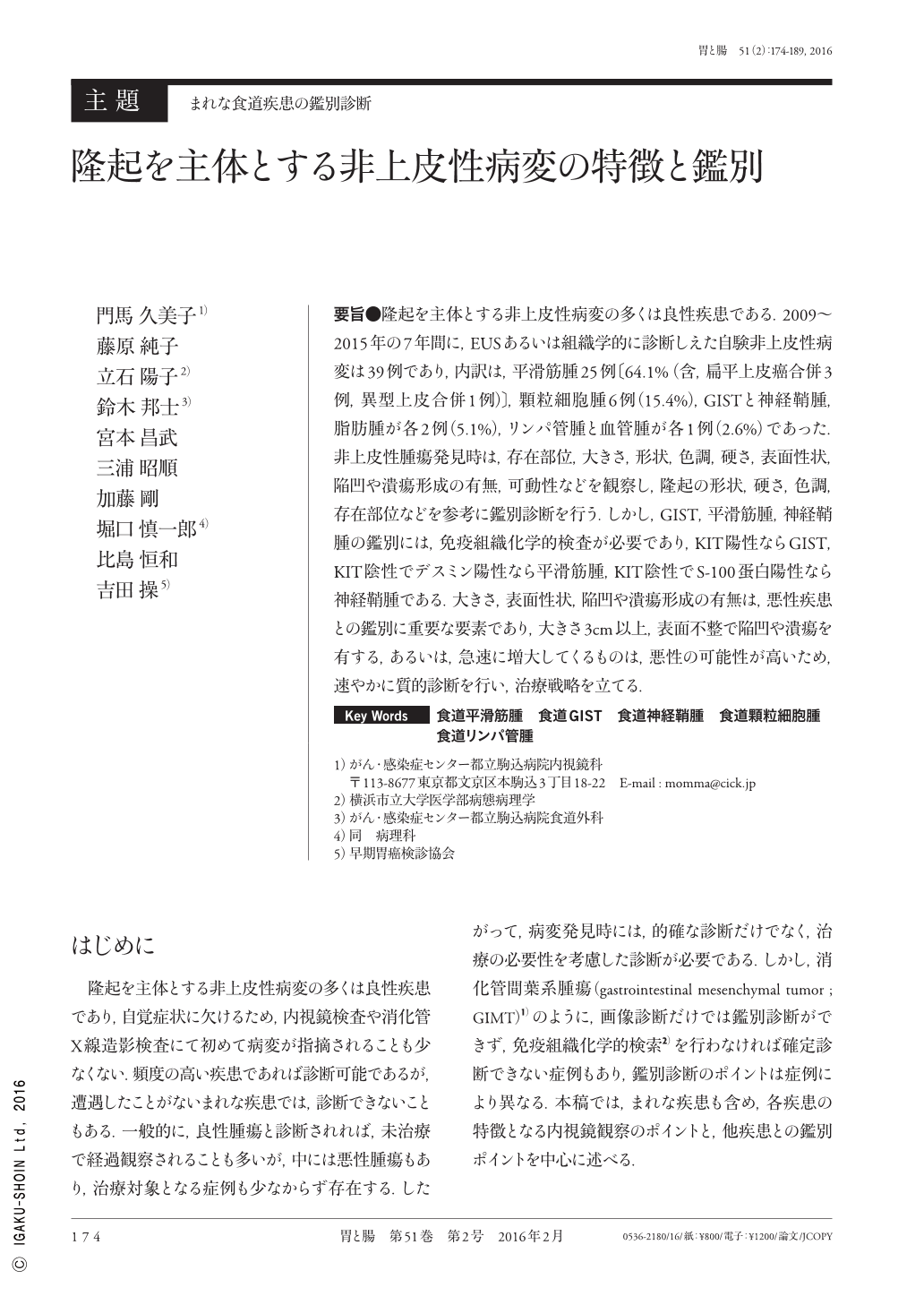
要旨●隆起を主体とする非上皮性病変の多くは良性疾患である.2009〜2015年の7年間に,EUSあるいは組織学的に診断しえた自験非上皮性病変は39例であり,内訳は,平滑筋腫25例〔64.1%(含,扁平上皮癌合併3例,異型上皮合併1例)〕,顆粒細胞腫6例(15.4%),GISTと神経鞘腫,脂肪腫が各2例(5.1%),リンパ管腫と血管腫が各1例(2.6%)であった.非上皮性腫瘍発見時は,存在部位,大きさ,形状,色調,硬さ,表面性状,陥凹や潰瘍形成の有無,可動性などを観察し,隆起の形状,硬さ,色調,存在部位などを参考に鑑別診断を行う.しかし,GIST,平滑筋腫,神経鞘腫の鑑別には,免疫組織化学的検査が必要であり,KIT陽性ならGIST,KIT陰性でデスミン陽性なら平滑筋腫,KIT陰性でS-100蛋白陽性なら神経鞘腫である.大きさ,表面性状,陥凹や潰瘍形成の有無は,悪性疾患との鑑別に重要な要素であり,大きさ3cm以上,表面不整で陥凹や潰瘍を有する,あるいは,急速に増大してくるものは,悪性の可能性が高いため,速やかに質的診断を行い,治療戦略を立てる.
Thirty-nine patients with elevated lesions of the esophagus with non-epithelial origin underwent endoscopy at our hospital from 2009 to 2015. A majority of these lesions were benign. The patients' final diagnoses were as follows:leiomyoma, 25 patients(64.1%); granular cell tumor, 6(15.4%); gastrointestinal stromal tumor(GIST), 2(5%); schwannoma, 2(5.1%); lipoma, 2(5.1%); lymphangioma, 1(2.6%); and hemangioma, 1(2.6%). In patients with leiomyoma, 3 patients had a combined case with squamous cell carcinoma confined to the mucosa and 1 patient with squamous intraepithelial neoplasia. Endoscopic differentiations were carried out based on endoscopic findings, such as the shape, solidity, color of the lesions, and the location in the esophagus. Immunohistochemical examinations were required for the differentiation of GIST, leiomyoma, and schwannoma. When the size of the lesions are>3cm or their shapes were irregular, relating to erosions or ulcerations, malignant changes should be considered. Rapidly growing tumors were also suggestive of malignancy. Prompt diagnoses and decision of treatments are required for patients with probable malignancy.

Copyright © 2016, Igaku-Shoin Ltd. All rights reserved.


