Japanese
English
- 有料閲覧
PPV購入案内
1,320円 (1,200円+税10%) こちらは、1785ページから1790ページの文献です。購入には医書.jp本会員登録が必要です。
会員登録完了後に改めて上記「購入サイトへ」を選択してください。
または「購入サイトへ」選択後に「購入ログイン」、「新規会員登録」の順に進んでください。
会員登録について詳しくはをご確認ください。
- Abstract 文献概要
- 1ページ目 Look Inside
- 参考文献 Reference
- サイト内被引用 Cited by
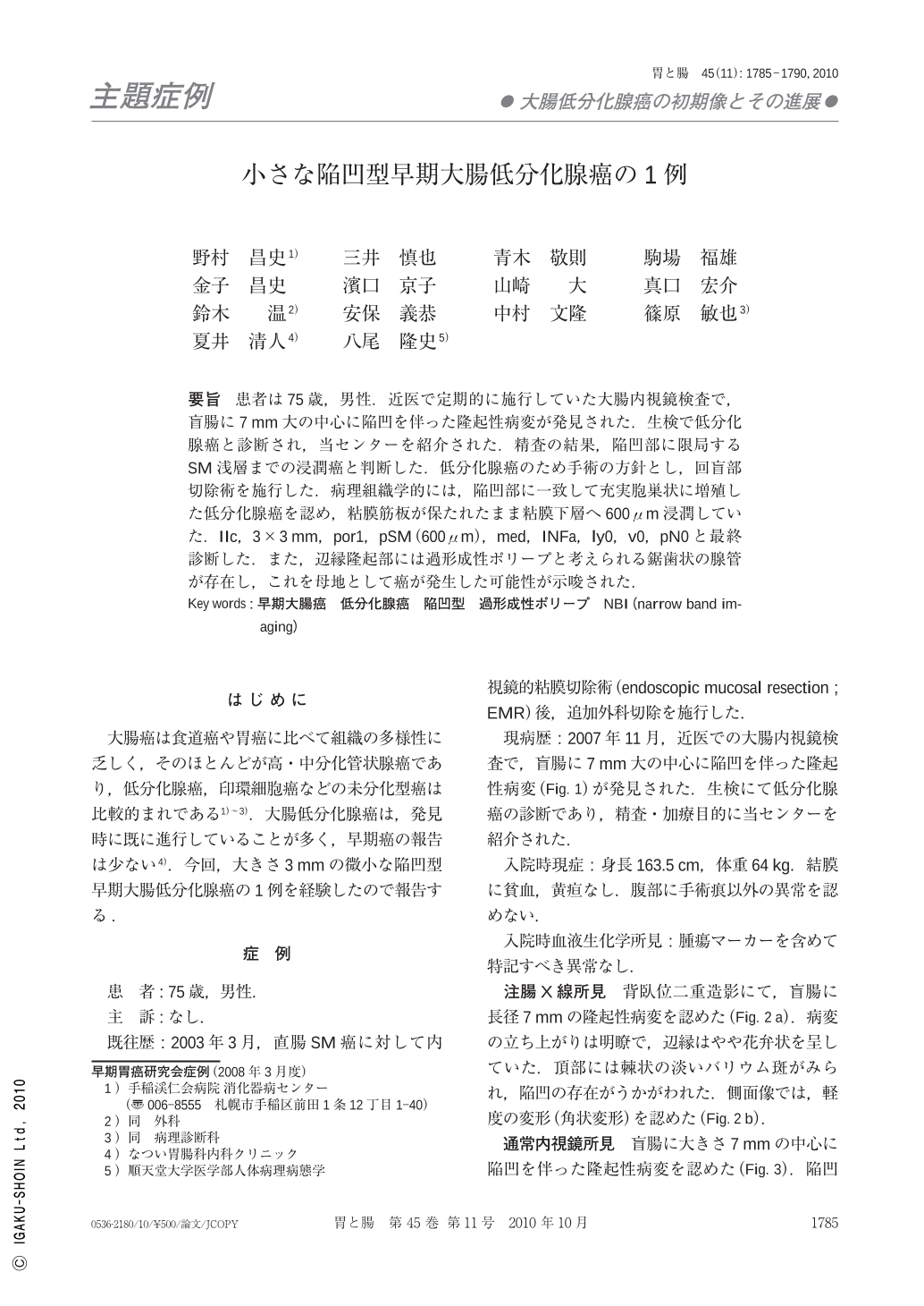
要旨 患者は75歳,男性.近医で定期的に施行していた大腸内視鏡検査で,盲腸に7mm大の中心に陥凹を伴った隆起性病変が発見された.生検で低分化腺癌と診断され,当センターを紹介された.精査の結果,陥凹部に限局するSM浅層までの浸潤癌と判断した.低分化腺癌のため手術の方針とし,回盲部切除術を施行した.病理組織学的には,陥凹部に一致して充実胞巣状に増殖した低分化腺癌を認め,粘膜筋板が保たれたまま粘膜下層へ600μm浸潤していた.IIc,3×3mm,por1,pSM(600μm),med,INFa,ly0,v0,pN0と最終診断した.また,辺縁隆起部には過形成性ポリープと考えられる鋸歯状の腺管が存在し,これを母地として癌が発生した可能性が示唆された.
A 75-year-old man was referred to our hospital because of poorly differentiated adenocarcinoma of the cecum detected by his primary doctor. Colonoscopic examination revealed a flat elevated lesion with a central depression, measuring 7mm in diameter in the cecum. This lesion was diagnosed as a submucosal invasive cancer occurring within a hyperplastic polyp. Ileocecal resection with regional lymph node dissection was thus performed. Histological findings showed poorly differentiated adenocarcinoma invading the submucosal layer(600μm)without lymphatic and venous invasion, and lymph node metastasis in a hyperplastic polyp. It is suggested that the development of this lesion from hyperplastic polyp to invasive cancer occurred via a serrated pathway.
Copyright © 2010, Igaku-Shoin Ltd. All rights reserved.