Japanese
English
- 有料閲覧
- Abstract 文献概要
- 1ページ目 Look Inside
- 参考文献 Reference
- サイト内被引用 Cited by
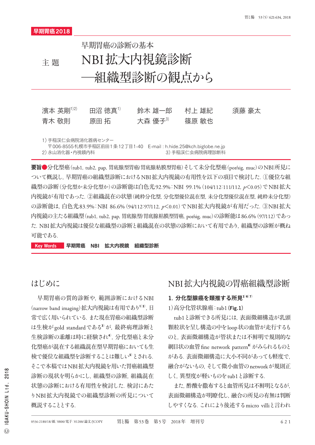
要旨●分化型癌(tub1,tub2,pap,胃底腺型胃癌/胃底腺粘膜型胃癌)そして未分化型癌(por/sig,muc)のNBI所見について概説し,早期胃癌の組織型診断におけるNBI拡大内視鏡の有用性を以下の項目で検討した.①優位な組織型の診断(分化型か未分化型か)の診断能は白色光92.9%:NBI 99.1%(104/112:111/112,p<0.05)でNBI拡大内視鏡が有用であった.②組織混在の状態(純粋分化型,分化型優位混在型,未分化型優位混在型,純粋未分化型)の診断能は,白色光83.9%:NBI 86.6%(94/112:97/112,p=0.663)であった.③NBI拡大内視鏡の主たる組織型(tub1,tub2,pap,胃底腺型/胃底腺粘膜型胃癌,por/sig,muc)の診断能は86.6%(97/112)であった.NBI拡大内視鏡は優位な組織型の診断において有用であり,組織型の診断が概ね可能である.
We investigated the capacity of NBI(narrow-band imaging)to detect differentiated cancers(tub1, tub2, pap, fundic gland-type gastric adenocarcinoma)and undifferentiated carcinomas(por/sig, muc), as well as its usefulness in the histological diagnosis of early gastric cancer. The following findings were obtained:(1)the diagnostic abilities of white light and NBI for dominant tissue type(differentiated or undifferentiated)were 92.6% and 99.1% respectively(104/112:111/112, p<0.05) ; (2)the diagnostic abilities of white light and NBI for mixed tissue conditions(purely differentiated, mostly differentiated, mostly undifferentiated, and purely undifferentiated)were 83.9% and 86.6%, respectively(94/112:97/112, p<0.01) ; and(3)the diagnostic ability of NBI for the main tissue types(tub1, tub2, fundic gland-type, fundic gland mucosa-type, pap, por/sig, muc), which are often targeted by this technique, was 86.6%(97/112). Taken together, these results demonstrate the usefulness of NBI magnifying endoscopy for diagnosing the status of both dominant and mixed tissue types and that tissue-type diagnosis was generally possible.

Copyright © 2018, Igaku-Shoin Ltd. All rights reserved.


