Japanese
English
- 有料閲覧
PPV購入案内
1,320円 (1,200円+税10%) こちらは、1514ページから1521ページの文献です。購入には医書.jp本会員登録が必要です。
会員登録完了後に改めて上記「購入サイトへ」を選択してください。
または「購入サイトへ」選択後に「購入ログイン」、「新規会員登録」の順に進んでください。
会員登録について詳しくはをご確認ください。
- Abstract 文献概要
- 1ページ目 Look Inside
- 参考文献 Reference
- サイト内被引用 Cited by
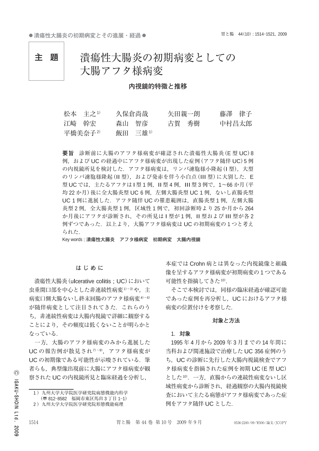
要旨 診断前に大腸のアフタ様病変が確認された潰瘍性大腸炎(E型UC)8例,およびUCの経過中にアフタ様病変が出現した症例(アフタ随伴UC)5例の内視鏡所見を検討した.アフタ様病変は,リンパ濾胞様小隆起(I型),大型のリンパ濾胞様隆起(II型),および発赤を伴う小白点(III型)に大別した.E型UCでは,主たるアフタはI型1例,II型4例,III型3例で,1~66か月(平均22か月)後に全大腸炎型UC6例,左側大腸炎型UC1例,ないし直腸炎型UC1例に進展した.アフタ随伴UCの罹患範囲は,直腸炎型1例,左側大腸炎型2例,全大腸炎型1例,区域性1例で,初回診断時より25か月から264か月後にアフタが診断され,その所見はI型が1例,II型およびIII型が各2例ずつであった.以上より,大腸アフタ様病変はUCの初期病変の1つと考えられた.
We compared colonoscopic findings in eight patients with early ulcerative colitis(E-UC)and five patients with UC accompanied by aphthous lesions(A-UC). One E-UC patient was affected by type I aphthous lesions(small protrusion with minimal depression), four E-UC patients by type II aphthous lesions(obvious protrusion with minimal depression), and three E-UC patients by type III aphthous lesions(white spot surrounded by reddish mucosa). During the subsequent periods ranging from 1 to 66 months, six E-UC patients progressed to full UC, one patient to left-sided colitis and the other patient to proctitis. The extent of UC in the A-UC group was the entire colon in one patient, the left colon in two patients, the rectum in one patient, and segmental involvement in one patient. The aphthous lesions in those patients were type I in one patient, type II in two patients, type III in two patients. These observations suggest that aphthous lesions are a precursor of typical involvement of the large intestine in UC.
Copyright © 2009, Igaku-Shoin Ltd. All rights reserved.