Japanese
English
- 有料閲覧
PPV購入案内
1,320円 (1,200円+税10%) こちらは、969ページから977ページの文献です。購入には医書.jp本会員登録が必要です。
会員登録完了後に改めて上記「購入サイトへ」を選択してください。
または「購入サイトへ」選択後に「購入ログイン」、「新規会員登録」の順に進んでください。
会員登録について詳しくはをご確認ください。
- Abstract 文献概要
- 1ページ目 Look Inside
- 参考文献 Reference
- サイト内被引用 Cited by
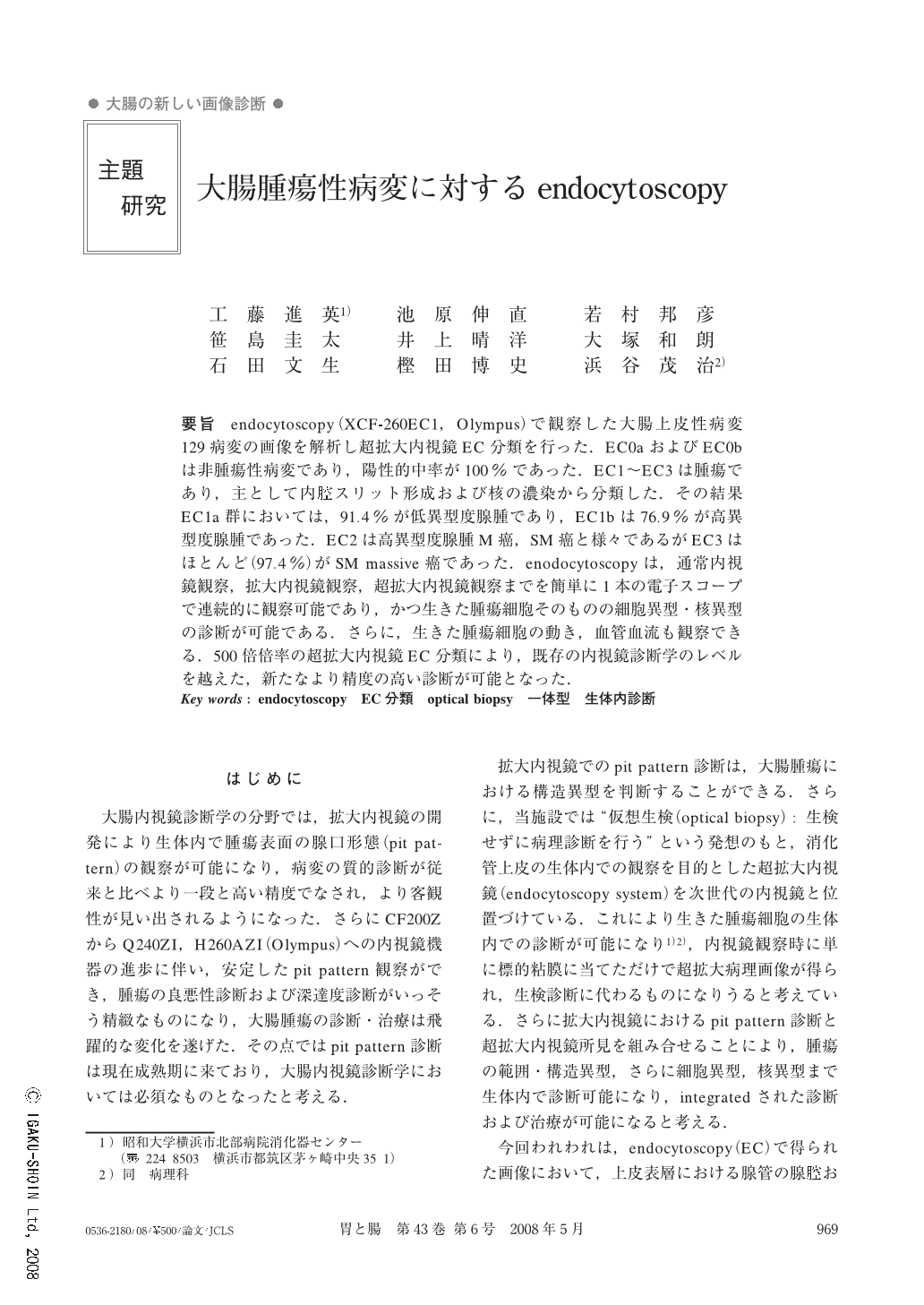
endocytoscopy(XCF-260EC1,Olympus)で観察した大腸上皮性病変129病変の画像を解析し超拡大内視鏡EC分類を行った.EC0aおよびEC0bは非腫瘍性病変であり,陽性的中率が100%であった.EC1~EC3は腫瘍であり,主として内腔スリット形成および核の濃染から分類した.その結果EC1a群においては,91.4%が低異型度腺腫であり,EC1bは76.9%が高異型度腺腫であった.EC2は高異型度腺腫M癌,SM癌と様々であるがEC3はほとんど(97.4%)がSM massive癌であった.enodocytoscopyは,通常内視鏡観察,拡大内視鏡観察,超拡大内視鏡観察までを簡単に1本の電子スコープで連続的に観察可能であり,かつ生きた腫瘍細胞そのものの細胞異型・核異型の診断が可能である.さらに,生きた腫瘍細胞の動き,血管血流も観察できる.500倍倍率の超拡大内視鏡EC分類により,既存の内視鏡診断学のレベルを越えた,新たなより精度の高い診断が可能となった.
In this study, we used the integrated type EC (XCF-260EC1, Olympus, Tokyo, Japan). We classified the EC findings into 6 groups and investigated whether the classification was useful for the differential diagnosis. The subjects were 129 lesions from May, 2005 to October, 2007 which were firstly detected by an ordinary view, then stained with 1% methylene blue, and observed, using the EC view. EC 0a: uniform glands with round lumen, EC 0b: uniform glands with serrated lumen, EC 1a: uniform glands with slit-like lumen and faintly stained fusiform nuclei, EC 1b: uniform glands with enlarged and darkly stained nuclei. EC 2: irregularly shaped glands with enlarged and distorted nuclei. EC 3: destroyed gland structure with enlarged and distorted nuclei. The final pathological diagnosis was normal mucosa in 5 cases, hyperplastic polyp in 6 cases, low grade adenoma in 39 cases, high grade adenoma or minimally invasive cancer in 34, massively invasive cancer in 45 cases. Positive predictive value of each EC group was as follows. EC 0 (non-tumor): 100%. EC 1a: (low grade adenoma): 91.4%. EC 2 (high grade adenoma or invasive cancer): 88.9%. EC 3(invasive cancer): 97.4%. The integrated type EC system enabled us to observe colorectal lesions at the cellular level in vivo. Our new classification of EC images corresponded well with the final pathological diagnosis. Endocytoscopy was especially useful for differential diagnosis between neoplastic and non-neoplastic lesions.
Copyright © 2008, Igaku-Shoin Ltd. All rights reserved.