Japanese
English
- 有料閲覧
PPV購入案内
1,320円 (1,200円+税10%) こちらは、581ページから588ページの文献です。購入には医書.jp本会員登録が必要です。
会員登録完了後に改めて上記「購入サイトへ」を選択してください。
または「購入サイトへ」選択後に「購入ログイン」、「新規会員登録」の順に進んでください。
会員登録について詳しくはをご確認ください。
- Abstract 文献概要
- 1ページ目 Look Inside
- 参考文献 Reference
- サイト内被引用 Cited by
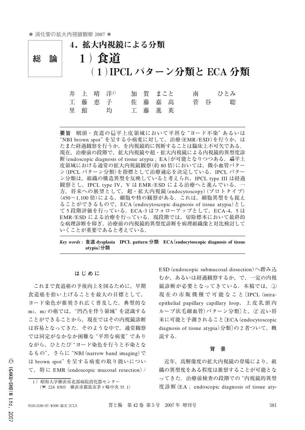
要旨 咽頭・食道の扁平上皮領域において平坦な“ヨード不染”あるいは“NBI brown spot”を呈する小病変に対して,治療(EMR/ESD)を行うか,はたまた経過観察を行うか,を内視鏡的に判断することは臨床上不可欠である.現在,治療前の段階で,拡大内視鏡や超・拡大内視鏡による内視鏡的異型度診断(endoscopic diagnosis of tissue atypia;EA)が可能となりつつある.扁平上皮領域における通常の拡大内視鏡観察(約80倍)においては,微小血管パターン(IPCLパターン分類)を指標として治療適応を決定している.IPCLパターン分類は,組織の構造異型を反映していると考えられ,IPCL type IIIは経過観察とし,IPCL type IV,VはEMR/ESDによる治療へと進んでいる.一方,将来への展望として,超・拡大内視鏡(endocytoscopy)(プロトタイプ)(450~1,100倍)による,細胞や核の観察がある.これは,細胞異型をも捉えることができるもので,ECA(endocytoscopic diagnosis of tissue atypia)として5段階評価を行っている.ECA-3はフォローアップとして,ECA-4,5はEMR/ESDによる治療を行っている.現段階では,切除標本において最終的な病理診断を仰ぎ,治療前の内視鏡的異型度診断を病理組織像と対比検討していくことが重要であると考えている.
Standard magnifying endoscopy has approximately 100-fold magnifying power. IPCL pattern is diagnosed with it. NBI (narrow band imaging) system is extremely useful to effectively recognize IPCL as a brown spot. In IPCL type classification, type I mainly includes normal epithelium. Type II corresponds to inflammatory change or non-neoplastic tissue and type III reflects border-line lesions. Type IV strongly suggests carcinoma in situ. Type V-1is definitely diagnosed as carcinoma in situ.
Endocytoscopy has approximately 500-fold magnification, which enables observation of cell and nucleus. Endocytoscopic images are classified into five categories from normal epithelium to malignant tissue as ECA classification (endocytoscopic atypism classification). ECA IV and V are considered to be treated in a clinical setting.
Copyright © 2007, Igaku-Shoin Ltd. All rights reserved.