Japanese
English
- 有料閲覧
PPV購入案内
1,320円 (1,200円+税10%) こちらは、727ページから732ページの文献です。購入には医書.jp本会員登録が必要です。
会員登録完了後に改めて上記「購入サイトへ」を選択してください。
または「購入サイトへ」選択後に「購入ログイン」、「新規会員登録」の順に進んでください。
会員登録について詳しくはをご確認ください。
- Abstract 文献概要
- 1ページ目 Look Inside
- 参考文献 Reference
- サイト内被引用 Cited by
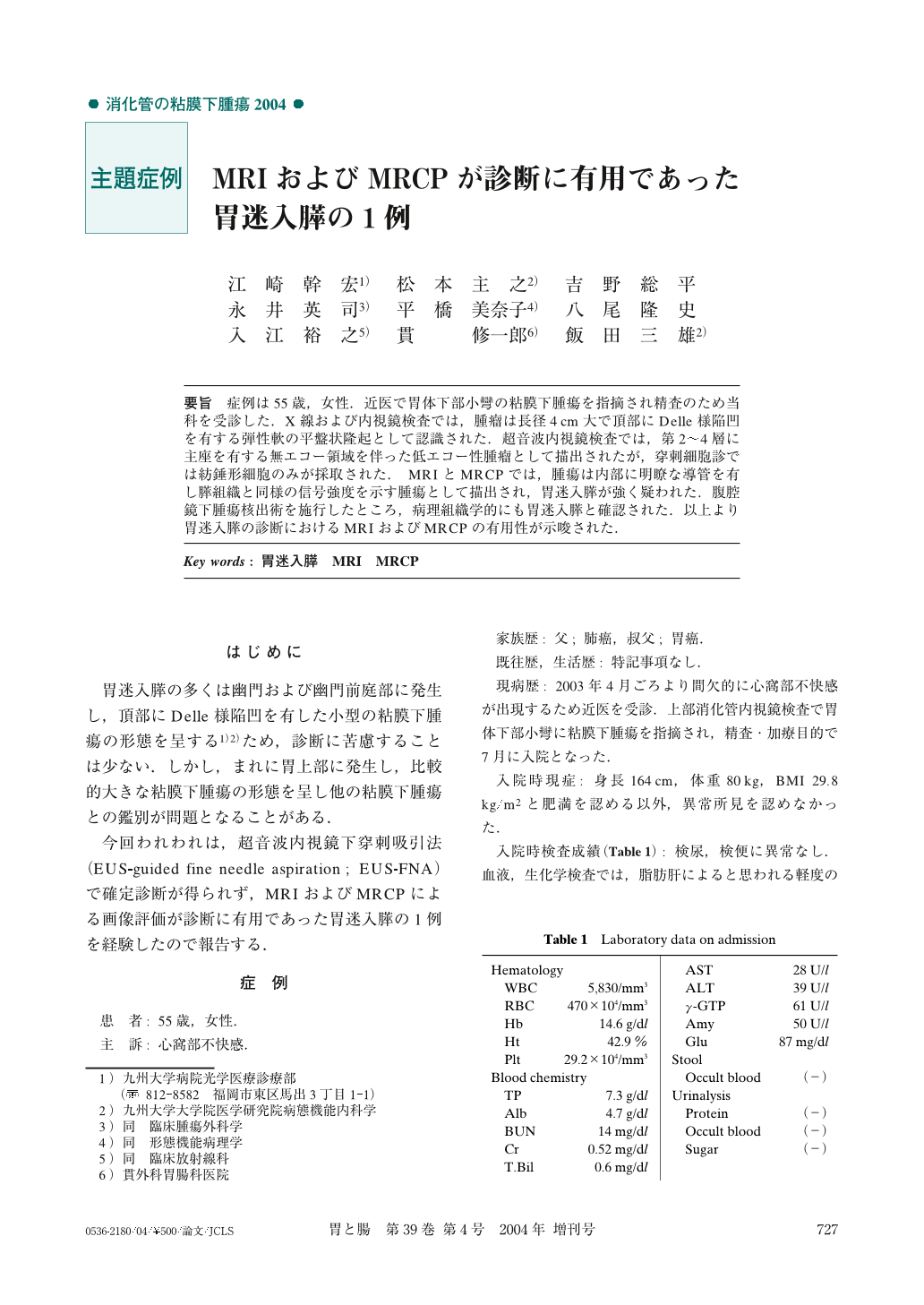
要旨 症例は55歳,女性.近医で胃体下部小彎の粘膜下腫瘍を指摘され精査のため当科を受診した.X線および内視鏡検査では,腫瘤は長径4cm大で頂部にDelle様陥凹を有する弾性軟の平盤状隆起として認識された.超音波内視鏡検査では,第2~4層に主座を有する無エコー領域を伴った低エコー性腫瘤として描出されたが,穿刺細胞診では紡錘形細胞のみが採取された.MRIとMRCPでは,腫瘍は内部に明瞭な導管を有し膵組織と同様の信号強度を示す腫瘍として描出され,胃迷入膵が強く疑われた.腹腔鏡下腫瘍核出術を施行したところ,病理組織学的にも胃迷入膵と確認された.以上より胃迷入膵の診断におけるMRIおよびMRCPの有用性が示唆された.
A 55-year-old female with a gastric tumor was referred to our institution. Under esophagogastroduodenoscopy, a sessile submucosal tumor with nodular surface and intact overlying mucosa was found on the lesser curvature of the gastric body. Radiography depicted the tumor as an elastic soft mass with central umbilication, measuring4cm in diameter. EUS showed the tumor to be a hypoechoic mass located mainly in the third and fourth layer of the gastric wall. A small anechoic area was also seen. Spindle-shaped cells without cellular atypia were obtained by EUS-guided fine-needle aspiration biopsy. Under MRCP, a dilated duct was depicted within the tumor. Based on these findings, we preoperatively diagnosed the tumor as a heterotopic pancreas of the stomach. The diagnosis was histologically verified in the resected specimen. It was thus suggested that MRCP may be a useful procedure for the diagnosis of heterotopic pancreas of the stomach.
1) Department of Endoscopic Diagnostics and Therapeutics, Kyushu University Hospital, Fukuoka, Japan
Copyright © 2004, Igaku-Shoin Ltd. All rights reserved.