Japanese
English
- 有料閲覧
- Abstract 文献概要
- 1ページ目 Look Inside
- 参考文献 Reference
- サイト内被引用 Cited by
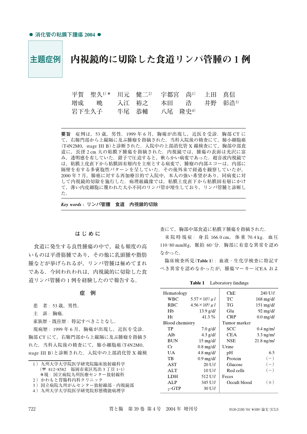
要旨 症例は,53歳,男性.1999年6月,胸痛が出現し,近医を受診.胸部CTにて,右肺門部から上縦隔に及ぶ腫瘤を指摘された.当科入院後の精査にて,肺小細胞癌(T4N2M0,stage III B)と診断された.入院中の上部消化管X線検査にて,胸部中部食道に,長径2cm大の粘膜下腫瘍を指摘された.内視鏡では,腫瘍の表面は光沢に富み,透明感を有していた.鉗子で圧迫すると,軟らかい病変であった.超音波内視鏡では,粘膜上皮直下から粘膜固有層内を主座とする病変で,腫瘤の内部エコーは,内部に隔壁を有する多嚢胞性パターンを呈していた.その後外来で経過を観察していたが,2000年7月,肺癌に対する再加療目的で入院中,本人の強い希望があり,同病変に対して内視鏡的切除を施行した.病理組織像では,粘膜上皮直下から粘膜固有層にかけて,薄い内皮細胞に覆われた大小不同のリンパ管が増生しており,リンパ管腫と診断した.
A 53-year-old man was referred to our hospital complaining of chest pain. The diagnosis of lung cancer was established by CT-guided mediastinal biopsy. When upper gastrointestinal examination was performed to rule out esophageal disease, a 2×1.2cm submucosal lesion in the middle third of the esophagus was demonstrated by chance. At endoscopy, this lesion was able to be recognized by the overlying normal mucosa, cystic translucency, and its deformation under pressure with biopsy forceps. Endoscopic ultrasonography revealed a multilocular cystic lesion derived from the submucosal layer. Endoscopic resection was performed, and the diagnosis was made histopathologically as lymphangioma.
1) Department of Clinical Radiology, Graduate School of Medical Sciences, Kyushu University, Fukuoka, Japan
2) Kawamoto Clinic, Fukuoka, Japan
3) National Kyushu Cancer Center, Fukuoka, Japan
4) Department of Anatomic Pathology, Graduate School of Medical Sciences, Kyushu University, Fukuoka, Japan

Copyright © 2004, Igaku-Shoin Ltd. All rights reserved.


