Japanese
English
- 有料閲覧
- Abstract 文献概要
- 1ページ目 Look Inside
- サイト内被引用 Cited by
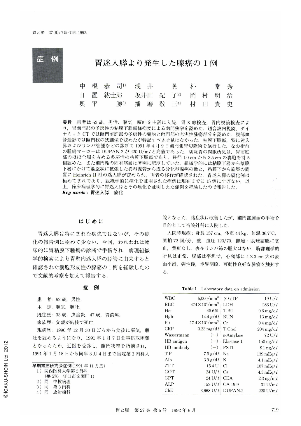
要旨 患者は62歳,男性.嘔気,嘔吐を主訴に入院.胃X線検査,胃内視鏡検査により,胃幽門部の多房性の粘膜下腫瘍様病変による幽門狭窄を認めた.超音波内視鏡,ダイナミックCTでは幽門前庭部の多房性の囊胞と幽門部の充実性腫瘍部分を認めた.腹部血管造影では幽門枝の狭細像を認めたが特記すべき所見はなかった.粘膜下腫瘍,特に迷入膵およびリンパ管腫などの診断で1991年4月9日幽門側胃切除術を施行した.なお術前の腫瘍マーカーはDUPAN-2が220U/mlと高値であった.切除胃の肉眼所見は,胃前庭部のほぼ全周を占める多房性の粘膜下腫瘍であり,長径1.0cmから3.5cmの囊胞を計5個認めた.また幽門輪の固有筋層は著明に肥厚していた.組織学的には粘膜下層から漿膜下層にかけて囊胞状に拡張した異型腺管から成る分化型腺癌の像と,粘膜下から筋層の間質にHeinrich Ⅱ型の迷入膵が認められ,両者の移行が確認された.胃迷入膵の癌化例は極めてまれであり,組織学的に癌化を証明された症例は現在までに15例にすぎない.以上,臨床病理学的に胃迷入膵とその癌化を証明しえた症例を経験したので報告した.
A 62-year-old male was admitted due to nausea and vomiting. On admission, physical examination showed no abnormalities, but blood analysis revealed an elevated DUPAN-2 tumor marker (220 U/ml). X-ray examination and endoscopic examination of the stomach showed a multilocular submucosal tumor at the pylorus with secondary pyloric stenosis. Ultrasonography and dynamic CT scan revealed polycystic lesion in the antrum and a solid mass at the pylorus. Abdominal angiography disclosed stenosis of the pyloric branch of the anterior superior pancreaticoduodenal artery but no other findings. A tentative diagnosis of a submucosal tumor in a heterotopic pancreas, and lymphangioma was made, and a distal gastrectomy was performed on April 9, 1991.
Macroscopic examination of the resected stomach showed multilocular submucosal tumors almost completely encircling the antrum with 5 cysts up to 4.0 cm in diameter. The propria muscle of the pyloric ring was markedly hypertrophied. Histologically, many cystic dilated ducts were scattered throughout the submucosa, down to the subserosa. These ducts were composed of stratified atypical mucin-secreting cuboidal cells invading stroma and perineurium consistent with welldifferentiated adenocarcinoma. In addition, scattered foci of Heinrich Ⅱ heterotopic pancreas were identified in the submucosa and the propria muscle. Transitional zones between ectopic pancreatic ductal tissue and the adenocarcinoma were identified.
Malignant transformation of the heterotopic pancreas in the stomach is very rare, and there are only 15 histologically proven cases. We report a histologically verifled case of a heterotopic pancreas with malignant transformation in the stomach.

Copyright © 1992, Igaku-Shoin Ltd. All rights reserved.


