Japanese
English
- 有料閲覧
PPV購入案内
1,320円 (1,200円+税10%) こちらは、706ページから711ページの文献です。購入には医書.jp本会員登録が必要です。
会員登録完了後に改めて上記「購入サイトへ」を選択してください。
または「購入サイトへ」選択後に「購入ログイン」、「新規会員登録」の順に進んでください。
会員登録について詳しくはをご確認ください。
- Abstract 文献概要
- 1ページ目 Look Inside
- 参考文献 Reference
- サイト内被引用 Cited by
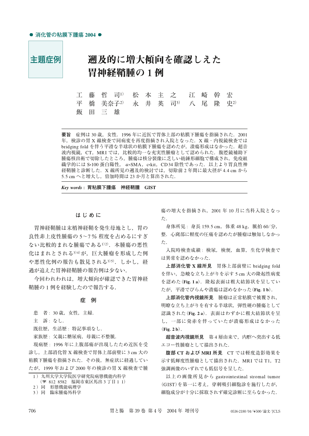
要旨 症例は30歳,女性.1996年に近医で胃体上部の粘膜下腫瘍を指摘された.2001年,検診の胃X線検査で同病変を再度指摘され入院となった.X線・内視鏡検査ではbridging foldを伴う平滑な半球状の粘膜下腫瘍を認めたが,潰瘍形成はなかった.超音波内視鏡,CT,MRIでは,比較的均一な充実性腫瘤として認められた.腹腔鏡補助下腫瘍核出術で切除したところ,腫瘍は核分裂像に乏しい紡錘形細胞で構成され,免疫組織学的にはS-100蛋白陽性,α-SMA,c-kit,CD34陰性であった.以上より胃良性神経鞘腫と診断した.X線所見の遡及的検討では,切除前2年間に最大径が4.4cmから5.5cmへと増大し,倍加時間は23か月と算出された.
A30-year-old female was referred to our institution, because of a gastric tumor which had increased in size during a 4-year period of observation. Radiography depicted a smooth semispherical tumor in the anterior wall of the upper gastric body. It measured 5.5cm in size. Under EGD, the tumor was observed as a sessile, submucosal tumor with intact overlying mucosa. EUS showed the tumor to be a hypoechoic mass in the fourth layer of the gastric wall. Because annual radiography showed the tumor to have increased in size, it was laparoscopically enuclulated. Microscopic examination revealed the tumor to be composed of spindle-shaped cells arranged in trabecular bundles without any mitotic figures. Lymphoid cuff was observed in the surface area of the tumor. Immunohistochemically, the tumor cells were positive for S-100 protein, but they were negative for c-kit, CD 34, α-SMA and desmin. Based on these findings, the diagnosis of benign gastric schwannoma was established.
1) Department of Medicine and Clinical Science, Graduate School of Medical Sciences, Kyushu University, Fukuoka, Japan
Copyright © 2004, Igaku-Shoin Ltd. All rights reserved.