Japanese
English
- 有料閲覧
PPV購入案内
1,320円 (1,200円+税10%) こちらは、99ページから109ページの文献です。購入には医書.jp本会員登録が必要です。
会員登録完了後に改めて上記「購入サイトへ」を選択してください。
または「購入サイトへ」選択後に「購入ログイン」、「新規会員登録」の順に進んでください。
会員登録について詳しくはをご確認ください。
- Abstract 文献概要
- 1ページ目 Look Inside
- 参考文献 Reference
- サイト内被引用 Cited by
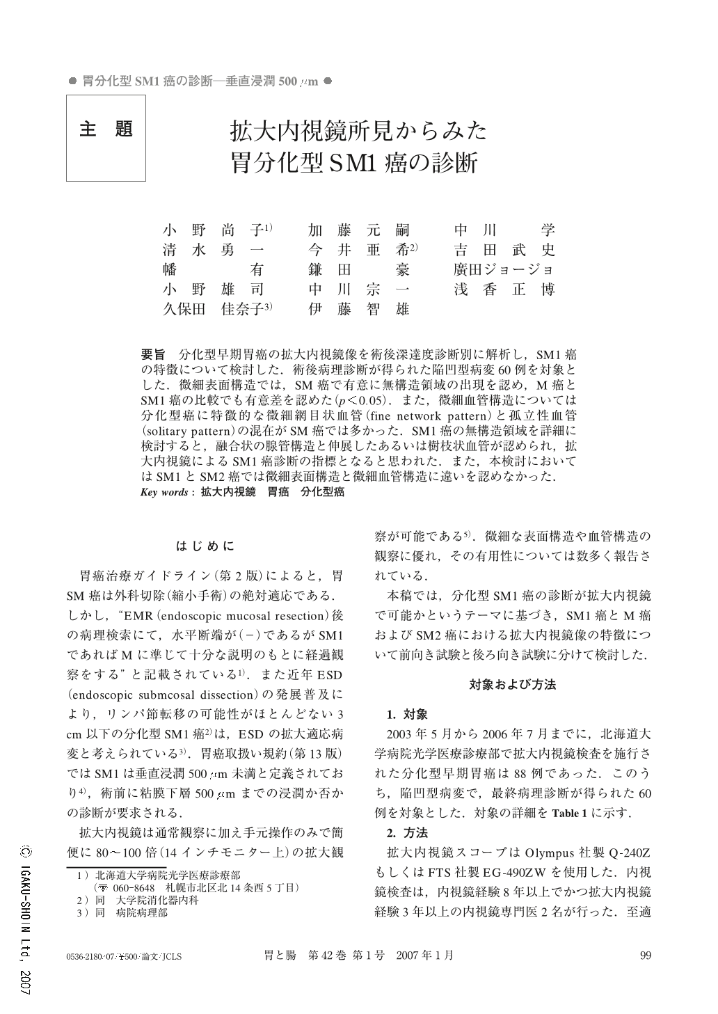
要旨 分化型早期胃癌の拡大内視鏡像を術後深達度診断別に解析し,SM1癌の特徴について検討した.術後病理診断が得られた陥凹型病変60例を対象とした.微細表面構造では,SM癌で有意に無構造領域の出現を認め,M癌とSM1癌の比較でも有意差を認めた(p<0.05).また,微細血管構造については分化型癌に特徴的な微細網目状血管(fine network pattern)と孤立性血管(solitary pattern)の混在がSM癌では多かった.SM1癌の無構造領域を詳細に検討すると,融合状の腺管構造と伸展したあるいは樹枝状血管が認められ,拡大内視鏡によるSM1癌診断の指標となると思われた.また,本検討においてはSM1とSM2癌では微細表面構造と微細血管構造に違いを認めなかった.
In order to determine the characteristics of SM1 invasive cancer, we analyzed magnifying endoscopic findings of intestinal-type early-stage gastric cancer according to postoperative diagnosis of invasion depth.
Sixty cases of depressed type early gastric cancer for which postoperative diagnosis was obtained were investigated. In the image of the surface microstructure, a nonstructural pattern was observed frequently in SM cancer, and there was a significant difference in frequencies of its appearance between m cancer and SM1 cancer (p<0.05). Moreover, in images of the microvascular structure, a combination of a fine network pattern and an isolated pattern was observed frequently in sm cancer. Detailed observation of the nonstructural pattern in SM1 cancer revealed a fused pattern of tubular structures and abnormal microvessels such as stretched type and branched type. These may be effective as an indicator by magnifying endoscopy of SM1 cancer. However, the results of this study revealed no obvious differences between SM1 cancer and SM2 cancer in surface microstructure or microvascular pattern.
Copyright © 2007, Igaku-Shoin Ltd. All rights reserved.