Japanese
English
- 有料閲覧
PPV購入案内
1,320円 (1,200円+税10%) こちらは、892ページから901ページの文献です。購入には医書.jp本会員登録が必要です。
会員登録完了後に改めて上記「購入サイトへ」を選択してください。
または「購入サイトへ」選択後に「購入ログイン」、「新規会員登録」の順に進んでください。
会員登録について詳しくはをご確認ください。
- Abstract 文献概要
- 1ページ目 Look Inside
- 参考文献 Reference
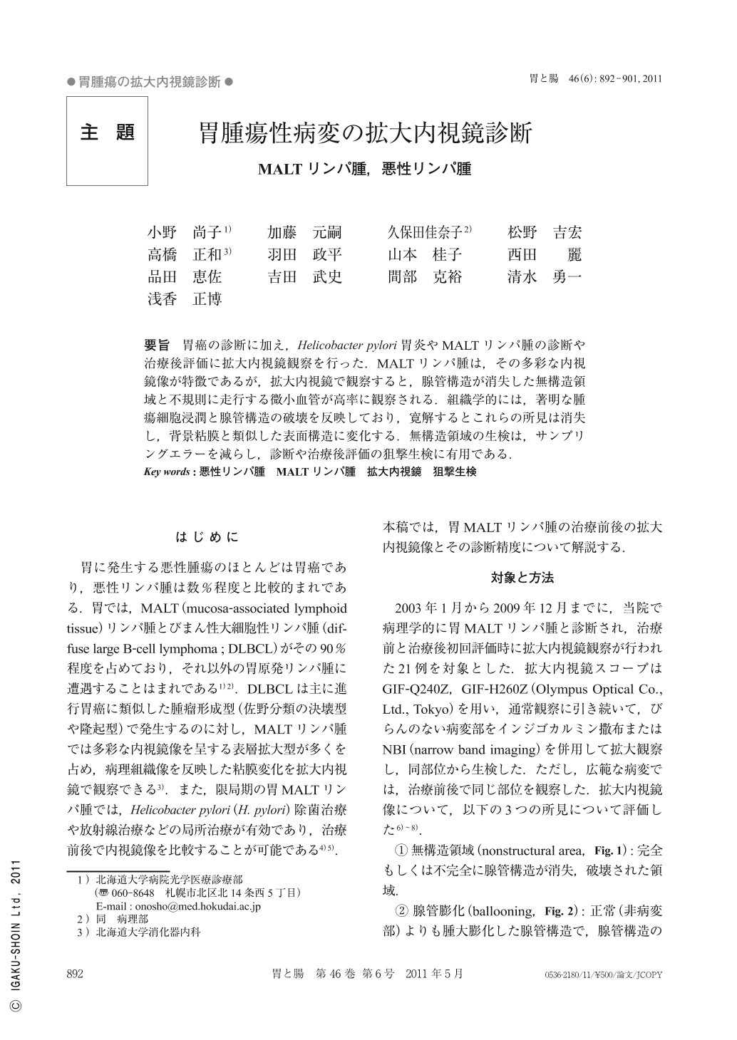
要旨 胃癌の診断に加え,Helicobacter pylori胃炎やMALTリンパ腫の診断や治療後評価に拡大内視鏡観察を行った.MALTリンパ腫は,その多彩な内視鏡像が特徴であるが,拡大内視鏡で観察すると,腺管構造が消失した無構造領域と不規則に走行する微小血管が高率に観察される.組織学的には,著明な腫瘍細胞浸潤と腺管構造の破壊を反映しており,寛解するとこれらの所見は消失し,背景粘膜と類似した表面構造に変化する.無構造領域の生検は,サンプリングエラーを減らし,診断や治療後評価の狙撃生検に有用である.
We used magnifying endoscopy for not only gastric cancer but also gastric MALT lymphoma and gastritis with Helicobacter pylori infection.
Although endoscopic appearances of MALT lymphoma are various, nonstructural areas without gastric glands and abnormal vessels with irregularity are characteristics in magnified endoscopic images. Magnified endoscopic findings reflect pathological views such as infiltration of lymphoma cells and destruction of gastric epithelium, and they change to similar surface microstructure in the background after eradication of lymphoma. Biopsy of the nonstructural area is useful to decrease sampling errors, to target biopsy for diagnosis and for evaluation after treatment.
Copyright © 2011, Igaku-Shoin Ltd. All rights reserved.