Japanese
English
特集 胸部の最新画像情報2024
[症例]
診断に苦慮した硬化性肺胞上皮腫の1例
A case of sclerosing pneumocytoma
佐藤 加奈子
1
,
河中 功一
2
,
田村 吉高
2
,
平井 俊範
2
Kanako Sato
1
1熊本赤十字病院 放射線科
2熊本大学大学院生命科学研究部 放射線診断学講座
1Department of Radiology Kumamoto Red-Cross Hospital
キーワード:
硬化性肺胞上皮腫
,
肺硬化性血管腫
,
肺腫瘍
Keyword:
硬化性肺胞上皮腫
,
肺硬化性血管腫
,
肺腫瘍
pp.83-88
発行日 2024年1月10日
Published Date 2024/1/10
DOI https://doi.org/10.18888/rp.0000002613
- 有料閲覧
- Abstract 文献概要
- 1ページ目 Look Inside
- 参考文献 Reference
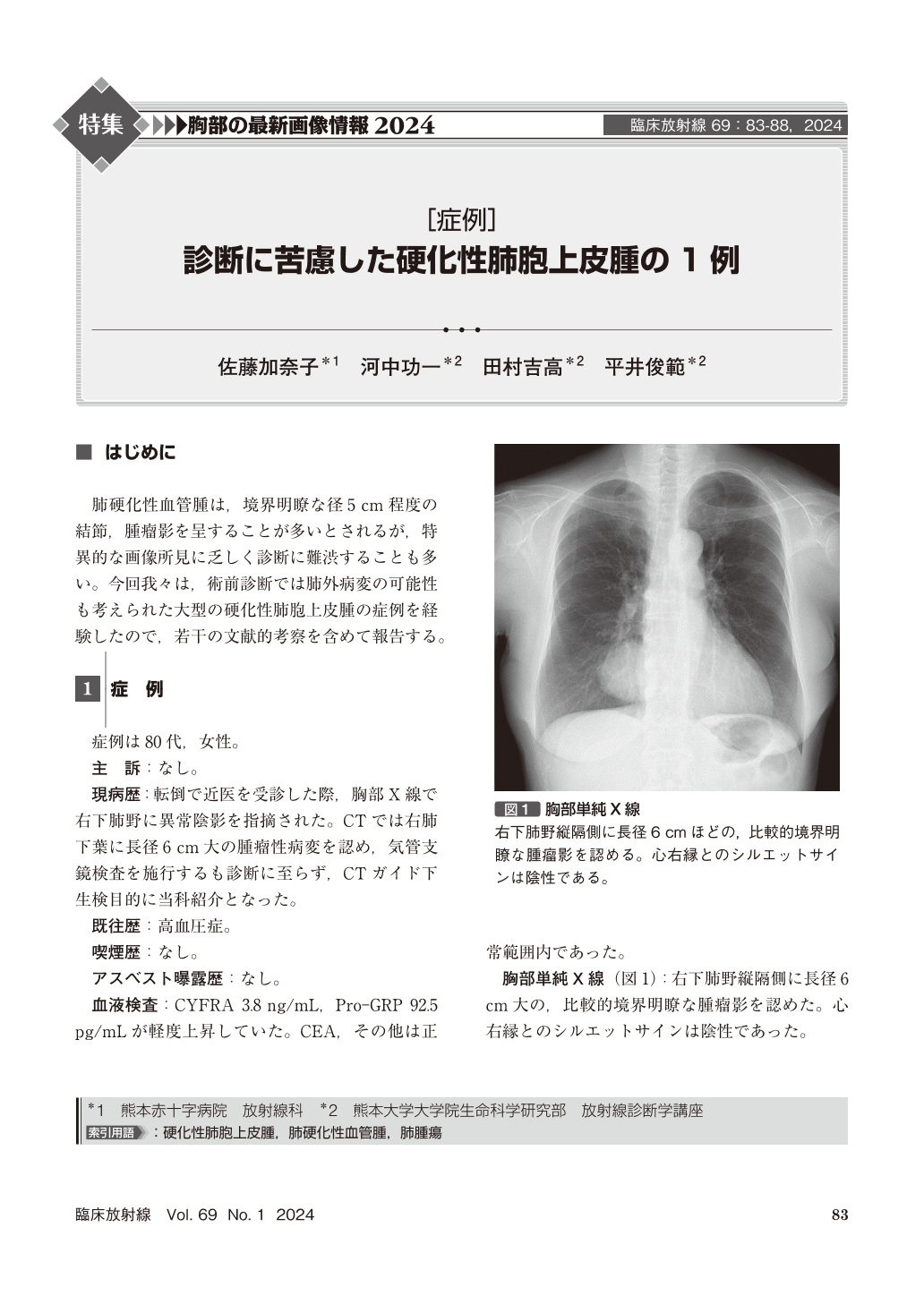
肺硬化性血管腫は,境界明瞭な径5cm程度の結節,腫瘤影を呈することが多いとされるが,特異的な画像所見に乏しく診断に難渋することも多い。今回我々は,術前診断では肺外病変の可能性も考えられた大型の硬化性肺胞上皮腫の症例を経験したので,若干の文献的考察を含めて報告する。
We report a case of sclerosing pneumocytoma of a woman in her 80s. Contrast-enhanced CT of the chest showed a large, slightly enhancing, well-defined mass in the right lower lobe. The mass appeared to be fed by the inferior phrenic artery and was suspected to be an extrapulmonary mass. The mass had diffusion restriction on MRI. As malignancy could not be ruled out, a right lower lobectomy was performed. After histopathological examination, the tumor was diagnosed as sclerosing pneumocytoma.

Copyright © 2024, KANEHARA SHUPPAN Co.LTD. All rights reserved.


