Japanese
English
- 有料閲覧
- Abstract 文献概要
- 1ページ目 Look Inside
- 参考文献 Reference
近年,胸部CTの普及と検出能力向上により,肺の微小病変の発見が増加している.それに伴い,肺病変に対してのCTガイド下生検を施行する機会も増加している.CTガイド下肺生検は末梢型肺病変の診断に対して有用な方法であるが,本検査にまれな合併症として空気塞栓症がある.われわれはCTガイド下生検施行後に冠状動脈空気塞栓を発症した症例を経験したので報告する.
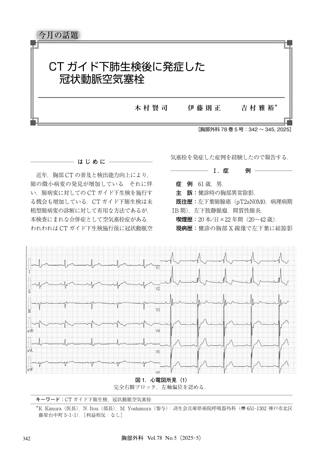
The patient was a 61-year-old man who was diagnosed with an abnormal chest shadow during a medical examination. A computed tomography (CT) scan revealed a nodular shadow in the left upper lobe, so a lung biopsy was performed for diagnostic purposes. Immediately after the biopsy, the patient experienced chest discomfort and a drop in pulse rate. An electrocardiogram showed ST elevation in Ⅱ, Ⅲ, aVf, and V1-V5. Chest CT revealed air in the right coronary artery and left ventricle, and the patient was diagnosed with air embolism. Morphine hydrochloride hydrate, nitroglycerin, and oxygen were administered. The chest pain improved the next morning. Chest CT performed the day after the examination showed that the air had disappeared. The patient’s condition improved, and he was discharged two days after the examination. Coronary artery air embolism occurred after the CT-guided biopsy, but he recovered without serious sequelae.
© Nankodo Co., Ltd., 2025