Japanese
English
- 販売していません
- Abstract 文献概要
- 1ページ目 Look Inside
- 参考文献 Reference
要旨
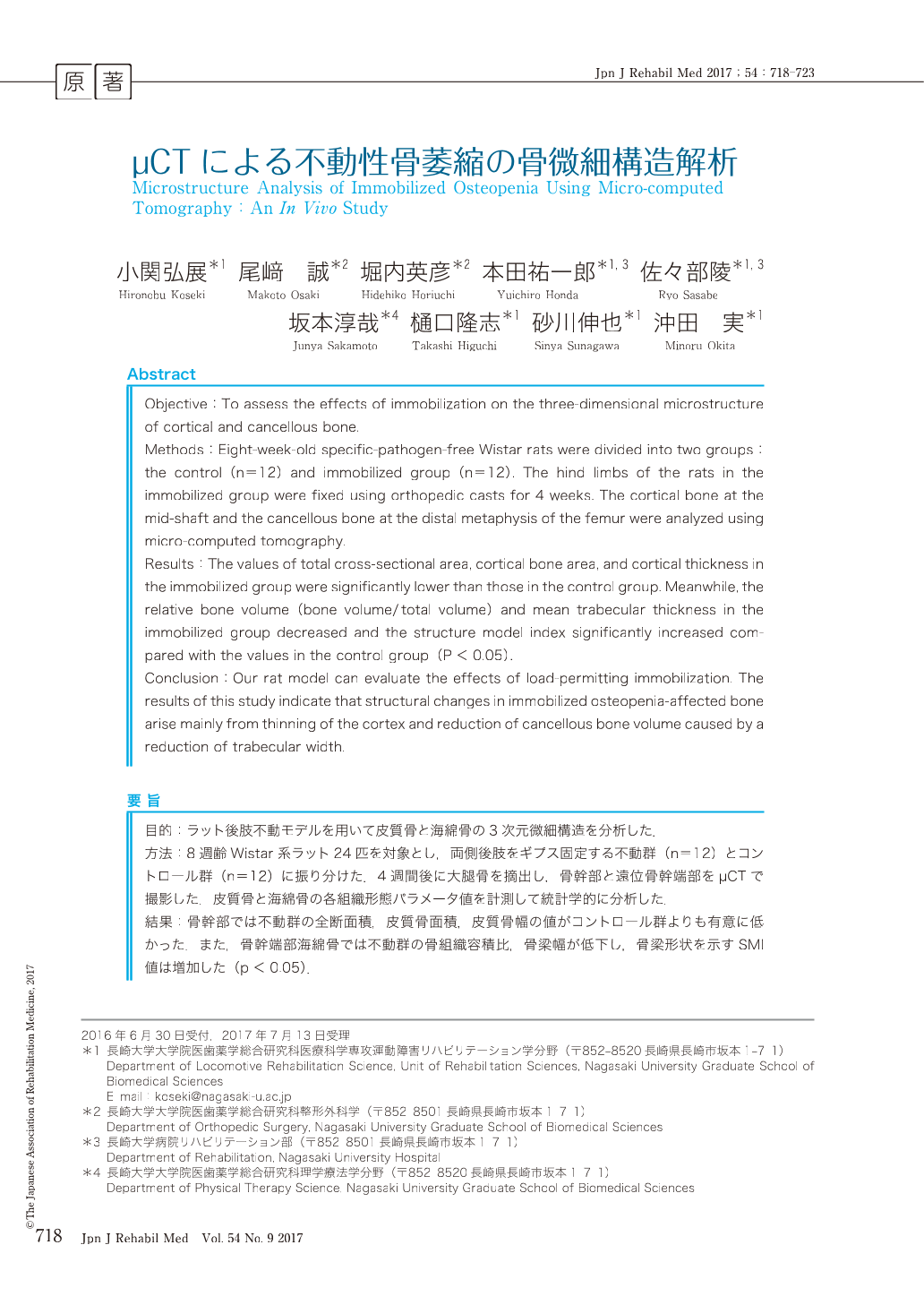
目的:ラット後肢不動モデルを用いて皮質骨と海綿骨の3次元微細構造を分析した.
方法:8週齢Wistar系ラット24匹を対象とし,両側後肢をギプス固定する不動群(n=12)とコントロール群(n=12)に振り分けた.4週間後に大腿骨を摘出し,骨幹部と遠位骨幹端部をµCTで撮影した.皮質骨と海綿骨の各組織形態パラメータ値を計測して統計学的に分析した.
結果:骨幹部では不動群の全断面積,皮質骨面積,皮質骨幅の値がコントロール群よりも有意に低かった.また,骨幹端部海綿骨では不動群の骨組織容積比,骨梁幅が低下し,骨梁形状を示すSMI値は増加した(p<0.05).
結論:本研究では「荷重を許容した不動化」が骨微細構造に与える影響を評価した.不動性骨萎縮の構造的特徴は,皮質骨幅の菲薄化と海綿骨の骨梁幅減少による骨容積比の低下である.
Objective:To assess the effects of immobilization on the three-dimensional microstructure of cortical and cancellous bone.
Methods:Eight-week-old specific-pathogen-free Wistar rats were divided into two groups:the control (n=12) and immobilized group (n=12). The hind limbs of the rats in the immobilized group were fixed using orthopedic casts for 4 weeks. The cortical bone at the mid-shaft and the cancellous bone at the distal metaphysis of the femur were analyzed using micro-computed tomography.
Results:The values of total cross-sectional area, cortical bone area, and cortical thickness in the immobilized group were significantly lower than those in the control group. Meanwhile, the relative bone volume (bone volume/total volume) and mean trabecular thickness in the immobilized group decreased and the structure model index significantly increased compared with the values in the control group (P<0.05).
Conclusion:Our rat model can evaluate the effects of load-permitting immobilization. The results of this study indicate that structural changes in immobilized osteopenia-affected bone arise mainly from thinning of the cortex and reduction of cancellous bone volume caused by a reduction of trabecular width.

Copyright © 2017, The Japanese Association of Rehabilitation Medicine. All rights reserved.


