Japanese
English
- 有料閲覧
- Abstract 文献概要
- 1ページ目 Look Inside
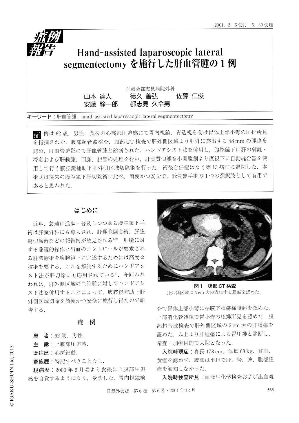
症例は62歳,男性.食後の心窩部圧迫感にて胃内視鏡,胃透視を受け胃体上部小彎の圧排所見を指摘された.腹部超音波検査,腹部CT検査で肝外側区域より肝外に突出する48mmの腫瘍を認め,肝血管造影にて肝血管腫と診断された.ハンドアシスト法を併用し,腹腔鏡下に肝の剥離・授動および肝動脈,門脈,胆管の処理を行い,肝実質切離を小開腹創より直視下に自動縫合器を使用して行う腹腔鏡補助下肝外側区域切除術を行った。術後合併症はなく第13病日に退院した.本術式は従来の腹腔鏡下肝切除術に比べ,簡便かつ安全で,低侵襲手術の1つの選択肢として有用であると思われた.
A 62-year old male presented with epigastric full sensation after meal. Endoscopic examination and barium meal study of the gastrointestinal tract showed impingement of the lesser curvature in the upper body of the stomach. An ultrasonography and computed tomography revealed a hepatic tumor measuring 48mm in size in the lateral segment. The tumor was diagnosed as hepatic hemangioma by celiac angiography. We performed hand-assisted laparoscopic lateral segmentectomy. Mobilization of the lateral segment and ligating vessels of the liver were done under laparoscopy.

Copyright © 2001, JAPAN SOCIETY FOR ENDOSCOPIC SURGERY All rights reserved.


