Japanese
English
- 有料閲覧
- Abstract 文献概要
- 1ページ目 Look Inside
- 参考文献 Reference
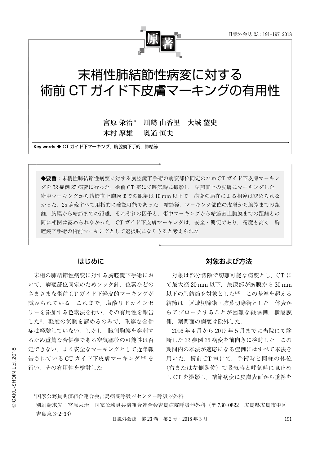
◆要旨:末梢性肺結節性病変に対する胸腔鏡下手術の病変部位同定のためCTガイド下皮膚マーキングを22症例25病変に行った.術前CT室にて呼気時に撮影し,結節直上の皮膚にマーキングした.術中マーキングから結節直上胸膜までの距離は10mm以下で,病変の局在による相違は認められなかった.25病変すべて用指的に確認可能であった.結節径,マーキング部位の皮膚から胸腔までの距離,胸膜から結節までの距離,それぞれの因子と,術中マーキングから結節直上胸膜までの距離との間に相関は認められなかった.CTガイド下皮膚マーキングは,安全・簡便であり,精度も高く,胸腔鏡下手術の術前マーキングとして選択肢になりうると考えられた.
While performing thoracoscopic wedge resection of the lung, the location of the lesion is generally identified by visual inspection or palpation. When difficulty in identification of the lesion by thoracoscopy is anticipated, preoperative marking is performed. However, complications and technical difficulties plague current marking techniques. To overcome this problem, we performed CT-guided skin marking above the pulmonary nodule. We used this technique to treat 25 lesions of 22 patients. Median tumor diameter was 9.5mm. All lesions were identified by finger tips via thoracoscopy, all nodules were constrained by ring forceps, and wedge resections were performed using a stapler. All lesions lay very close to the markings, as judged by finger palpation. No complications were encountered. The advantages of our technique are that it is simple and easy, air emboli are not an issue, the skin marking is rapid, and safety is assured.

Copyright © 2018, JAPAN SOCIETY FOR ENDOSCOPIC SURGERY All rights reserved.


