Japanese
English
- 有料閲覧
- Abstract 文献概要
- 1ページ目 Look Inside
I.はじめに
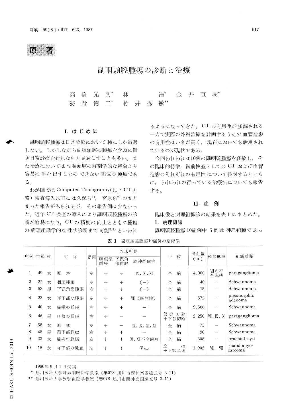
副咽頭腔腫瘍は日常診療において稀にしか遭遇しない。しかしながら副咽頭腔の腫瘍を念頭に置き日常診療を行わないと見過ごすことも多い。また治療においては副咽頭腔の解剖学的な特徴より容易に手を出すことのできない部位の腫瘍である。
わが国ではComputcd Tomography(以下CTと略)検査導入以前には久保ら1),宮原ら2)のまとまった報告がみられるが,その報告例は少なかった。近年CT検査の導入により副咽頭腔腫瘍の診断が容易になり,CTの精度の向上とともに腫瘍の病理組織学的な性状診断まで可能3,4)といわれるようになってきた。CTの有用性が強調される一方で実際の外科治療を計画するうえで血管造影の有用性はいまだ高く,現在においても活用されているのが現状である。
Ten cases of parapharyngeal space tumors are presented with discussions of the pathology, clinical aspects and treatment of the lesions in the parapharyngeal space. Use of computed tomographic (CT) scan and angiography on such lesions are discussed as they apply to management.
The most useful radiologic investigation is CT scan, but carotid angiography is still needed for the preoperative examination for these lesions.
The external cervical approach was applied in all cases. The surgical approach to skull base was reported.

Copyright © 1987, Igaku-Shoin Ltd. All rights reserved.


