Japanese
English
- 有料閲覧
- Abstract 文献概要
- 1ページ目 Look Inside
I.はじめに
頭頸部悪性腫瘍には一般に浅側頭動脈より逆行性に挿入した留置動注チューブからの化学療法と放射線照射を術前治療として行っている。この動注チューブの動脈内での先端の位置確認は,薬剤の効果的な投与のみならず重篤な合併症を防止すためにも重要である。それゆえに化学療法を開始するのに先立ってチューブ先端の位置確認は充分に検討しておかねばならない。従来この動注チューブ先端の位置確認は動注チューブより1%インジゴカルミン注入により染色される皮膚,粘膜の支配動脈より推測する色素注入法が一般に用いられている。この方法では動注チューブ先端が染色された部位より中枢に存在することを示すにすぎず確実なチューブ先端の同定は困難である。
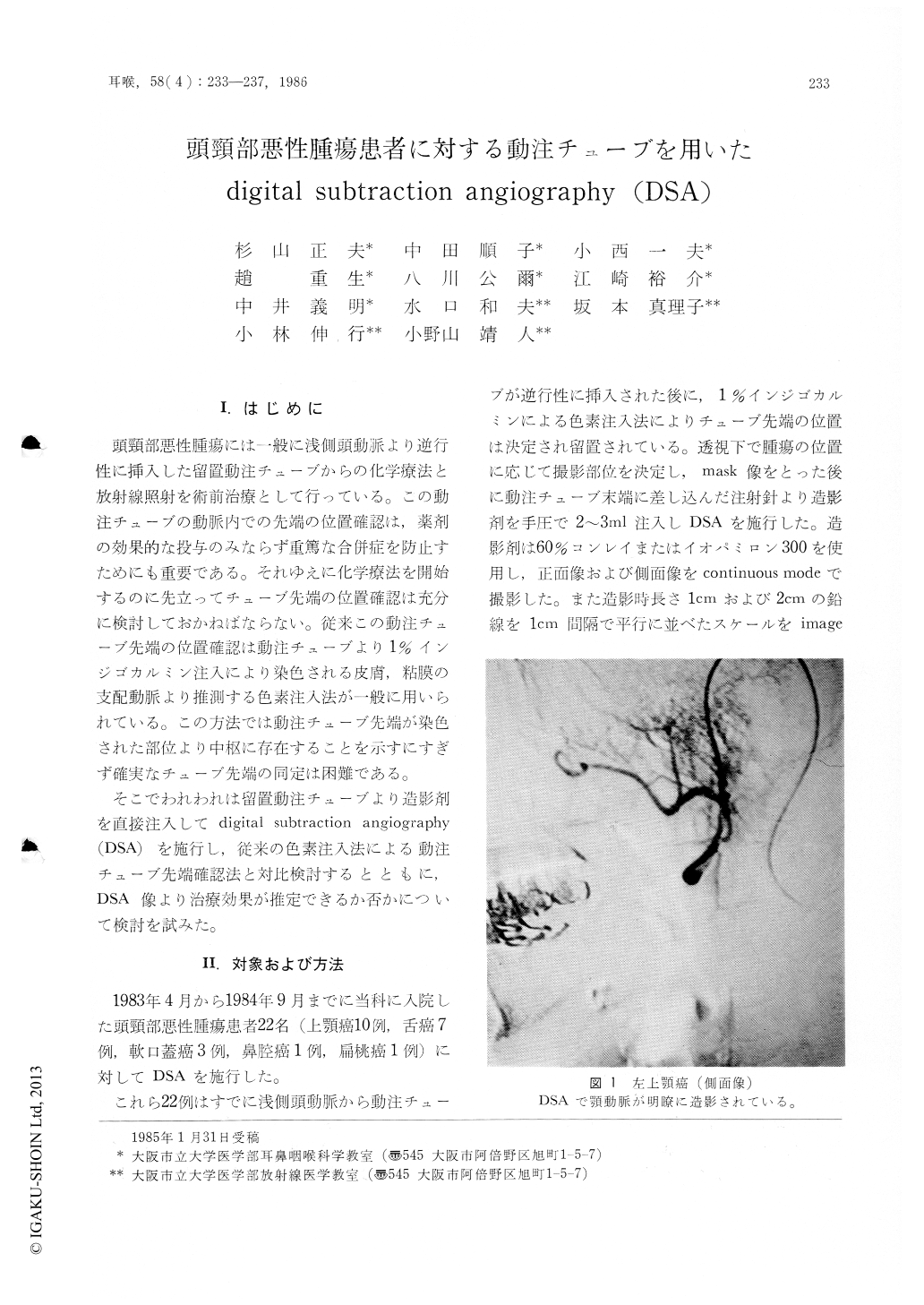
そこでわれわれは留置動注チューブより造影剤を直接注入してdigital subtraction angiography(DSA)を施行し,従来の色素注入法による動注チューブ先端確認法と対比検討するとともに,DSA像より治療効果が推定できるか否かについて検討を試みた。
Digital subtraction angiography (DSA) was performed through an indwelling catheter inserted into the superficial temporal artery on 22 cases of head and neck cancer. By means of DSA, the tip of the intraarterial catheter can be securely fixed in a short time without giving pain to the patient. In the 22 cases the tip of the catheter was adequately positioned by the conventional indigocarmine dye method. However, it was disclosed by DSA that the catheter was inserted deeper than expected in 4 of them. When observed under DSA, in such cases, the deeply inserted catheter could be easily adjusted to proper position.
Applications of DSA for estimation of the extent of tumor and evaluation of therapeutic effect were possible in certain cases, but satisfactory results could not be obtained hitherto.

Copyright © 1986, Igaku-Shoin Ltd. All rights reserved.


