Japanese
English
- 有料閲覧
- Abstract 文献概要
- 1ページ目 Look Inside
I.緒言
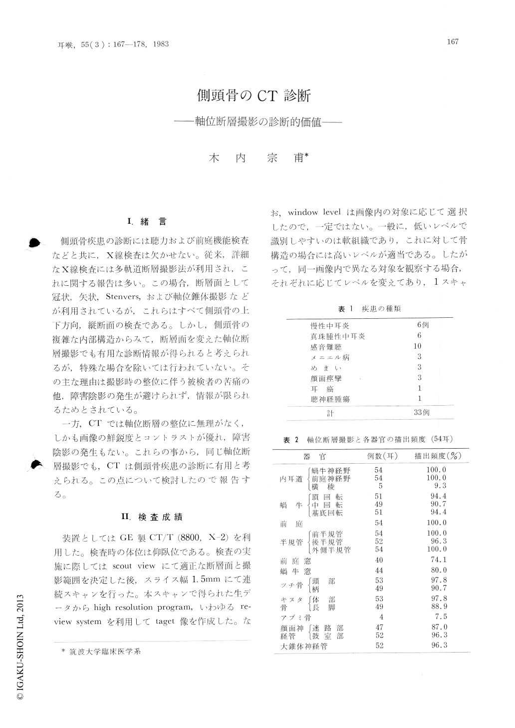
側頭骨疾患の診断には聴力および前庭機能検査などと共に,X線検査は欠かせない。従来,詳細なX線検査には多軌道断層撮影法が利用され,これに関する報告は多い。この場合、断層面として冠状,矢状,Stenvers,および軸位錐体撮影などが利用されているが,これらはすべて側頭骨の上下方向,縦断面の検査である。しかし,側頭骨の複雑な内部構造からみて,断層面を変えた軸位断層撮影でも有用な診断情報が得られると考えられるが,特殊な場合を除いては行われていない。その社な理由は撮影時の整位に伴う被検者の苦痛の他,障害陰影の発生が避けられず,情報が限られるためとされている。
一方,CTでは軸位断層の整位に無理がなく,しかも両像の鮮鋭度とコントラストが優れ,障害陰影の発生もない。これらの事から,同じ軸位断層撮影でも,CTは側頭骨疾患の診断に有用と考えられる。この点について検討したので報告する。
Axial CT scan was used to investigate the radiological details of the temporal bone of 33 patients with chronic otitis media, secondary cholesteatoma, sensorineural hearing loss, Meniere disease, vertigo, facial spasm, and neoplasma.
The axial scans showed anatomic details of the temporal bone, and at the same time clearly demonstrated the extent of the soft-tissue masses in the middle ears, as well as the destructions of the ossicles. Bone changes of the anterior walls of the epitympanum and external auditory meatus were more clearly demonstrated than by coronary CT scan. However, the axial scan had the disadvantages in demonstrating the stapes, crista transversa, and the mastoid portion of the facial canal.

Copyright © 1983, Igaku-Shoin Ltd. All rights reserved.


