Japanese
English
- 有料閲覧
- Abstract 文献概要
- 1ページ目 Look Inside
I.はじめに
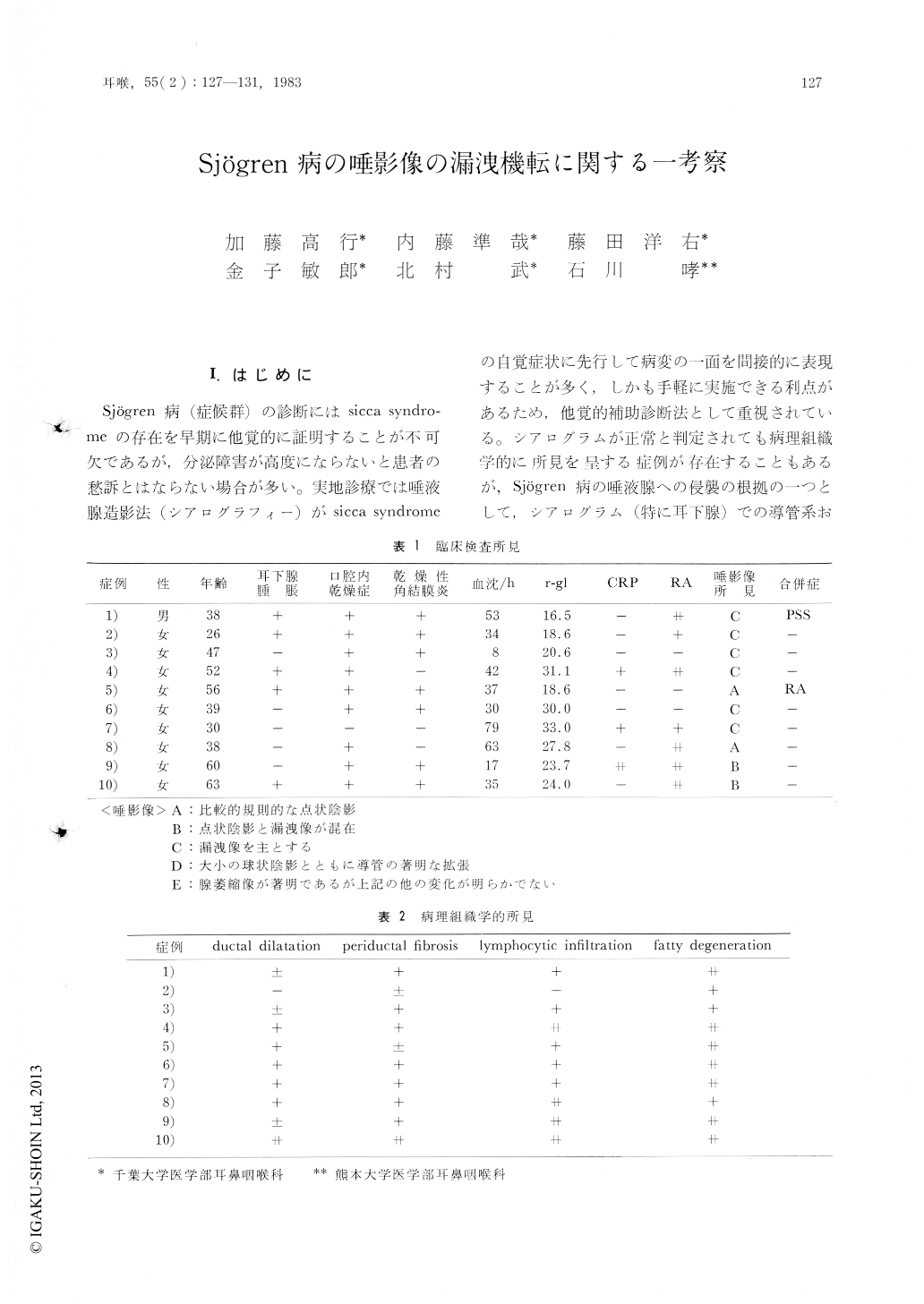
Sjögren病(症候群)の診断にはsicca syndromeの存在を早期に他覚的に証明することが不可欠であるが,分泌障害が高度にならないと患者の愁訴とはならない場合が多い。実地診療では唾液腺造影法(シアログラフィー)がsicca syndromeの自覚症状に先行して病変の一面を間接的に表現することが多く,しかも手軽に実施できる利点があるため,他覚的補助診断法として重視されている。シアログラムが正常と判定されても病理組織学的に所見を呈する症例が存在することもあるが,Sjögren病の唾液腺への侵襲の根拠の一つとして,シアログラム(特に耳下腺)での導管系および腺部の漏洩が挙げられる。Shearn1)のSjögren病の診断基準以来,唾液腺生検が確定診断の決め手として強調される傾向にあるが,われわれは唾液腺(耳下腺)生検と施行し碓診を得た症例をもとに,漏洩像の発生機転に関して病理組織学的に検討を加えたので,報告する。
Sialography is one of the useful procedures for the diagnosis of Sjögren's disease, because it reflects faithfully the histopathological processes of this disease.
The sialogram of Sjögren's disease is characterrized by punctate, globular, cavitary sialectasis due to acinar atrophy, fragmentation and disintegration of these structures, and thin or few ductulus due to myoepithelial hyperplasia which obliterates the lumen. In addition, the extravasation of contrast media from the larger ducts into surrounding tissue is another principal feature. To analyse the mechanism of extravasation of contrast media, the histological examination was undertaken in some cases to investigate elastic fiber around the larger ducts. In the end stage of Sjögren's disease, the disappearance or interruption of the elastic fiber were strikingly observed. This fact means that one of the causes of extravasation of contrast media is due to fragility of the ductal walls.

Copyright © 1983, Igaku-Shoin Ltd. All rights reserved.


