Japanese
English
- 有料閲覧
- Abstract 文献概要
- 1ページ目 Look Inside
Ⅰ.はじめに
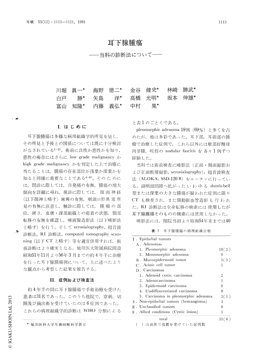
耳下腺腫瘍は多様な病理組織学的所見を呈し,その所見と予後との関係については既に十分検討がなされている1〜3)。術前に良性か悪性かを知り,悪性の場合にはさらにlow grade malignancyかhigh grade malignancyかを判定した上で治療に当たることは,腫瘍の存在部位が浅葉か深葉かを知ると同様に重要なことである4〜6)。そのためには,問診に際しては,自発痛の有無,腫瘍の増大傾向を詳細に尋ね,視診に際しては,顔面神経(以下顔神と略す)麻痺の有無,咽頭口腔異常所見の有無に注意し,触診に際しては,腫瘤の部位,硬さ,皮膚・深部組織との癒着の状態,頸部転移の有無を確認し,唾液腺造影法(以下唾影法と略す)を行う。そしてxerosialography,超音波診断法,RI診断法,computed tomography scanning(以下CTと略す)等を適宜併用すれば,術前診断はより確実となる。旭川医大附属病院開設昭和51年11月より56年3月までの約4年半に治療を行った耳下腺腫瘍例について,上に述べたような観点から考察した結果を報告する。
The preoperative diagnosis of the parotid gland tumors in 31 patients was evaluated. The results were as follows ; leakage of contrast media in sialogram of the parotid gland usually suggests malignant tumor. But, a few histologically proven benign tumors showed also leakage, so the relationships among the duct, capsule and parenchym were reexamined from the extirpated specimens. The ultrasonic examination was very useful for the differential diagnosis between benign and malignant tumor. But the standardization of this procedure requires some experiences. The repeated sialograms after the first trial might have shown leakage in malignant tumor, but no leakage in benign ones. So, this procedure is evaluated to be useful in the differential diagnosis.

Copyright © 1981, Igaku-Shoin Ltd. All rights reserved.


